
Submitted by jer5150 on Sun, 04/21/2013 - 13:30
Jason is taking a break (everyone needs one now and then). April's ECG will appear through May, as well. It's a good one!
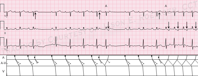
These are 3 simultaneous rhythm tracings that were once part of a full 12-lead ECG. Unfortunately, this ECG dates from a time period when our facility's computer database did not store complete 12-lead ECGs. The technology at the time was known as "reduced data", so much of the entire 12-lead ECG was deleted and only leads II, V1, and V5 were preserved for review by the cardiologist. This was in an attempt to reduce the amount of information that the computer would need to store over time. I can only imagine that this method was eventually deemed to be an undesirable way of storing ECG data. Later generations of computer storage would download the 12-lead ECG in its entirety.
All of that being said, what is this showing? Obviously, many things can not be determined without the remaining leads (e.g., QRS axis, chamber enlargement, etc) but the arrhythmic mechanism can still be determined.
Rate this content:
-

- jer5150's blog
- Log in or register to post comments
All our content is FREE & COPYRIGHT FREE for non-commercial use
Please be courteous and leave any watermark or author attribution on content you reproduce.
Comments
My guess:
My guess:
1st degree AV-block
Pauses due to non-conducted PAC's
retrograde P-waves at the end suggest AVRT???
my suspection
I think that there are atrial echoes + potential for Wenckebach involved - again..
Two cycles of wenckebach periods, both interupted by atrial echo.
And in the last cycle, the atrial echo starts run of AVNRT? Looking forward to the explanation :)!
2 nd degree type 2
2 nd degree type 2
Fast & Slow Pathways with Retrograde Adventure
Gorgeous tracing - and I'll look forward to Jason's laddergram! I see the following:
Delightful!
Ken Grauer, MD www.kg-ekgpress.com [email protected]
INTERPRETATION
Not much to add to Ken’s interpretation. As Ken points out, it’s the subtle changes of duration in the “coupling” interval that determines whether or not a PAC conducts. The longer post-ectopic cycles end with sinus beats (4th and 9th beats) that conduct with somewhat shorter P-R intervals implying that there is undoubtedly a potential for Wenckebach conduction at a faster sinus rate. The isolated retrograde P’-wave, following the 7th beat, can also be called an atrial “echo” or reversed reciprocal beat. The two conducted PACs are labled “A”. The upward-pointing arrows in V1 are marking the early ectopic atrial P’-waves whereas the downward-pointed arrows in lead II are marking the retrograde P’-waves.I am a stark proponent of using the term “nonconducted PAC” in place of the all-too-often confusing term “blocked PAC”. On more than one occasion, I have seen a patient get erroneously admitted for having a “new onset of A-V block” because the provider interpreting their ECG equated “blocked PAC” with being a type or form of A-V block. Invariably, healthcare providers hear the word “block” and reflexively think of heart block.
The reviewing cardiologist diagnosed this ECG simply as:
Normal sinus rhythm 2nd degree AV block (Mobitz i, Wenckebach)
There are clearly no “dropped” beats.
Jason E. Roediger - Certified Cardiographic Technician (CCT)
[email protected]
Dual AV node conduction
I presume we can say the dual AV node conduction is initiated by the PAC?
Ryan
You presume correctly
Yes Ryan, I think this would count as dual A-V nodal conduction.
Jason E. Roediger - Certified Cardiographic Technician (CCT)
[email protected]