

Submitted by Dawn on Wed, 05/09/2012 - 18:19
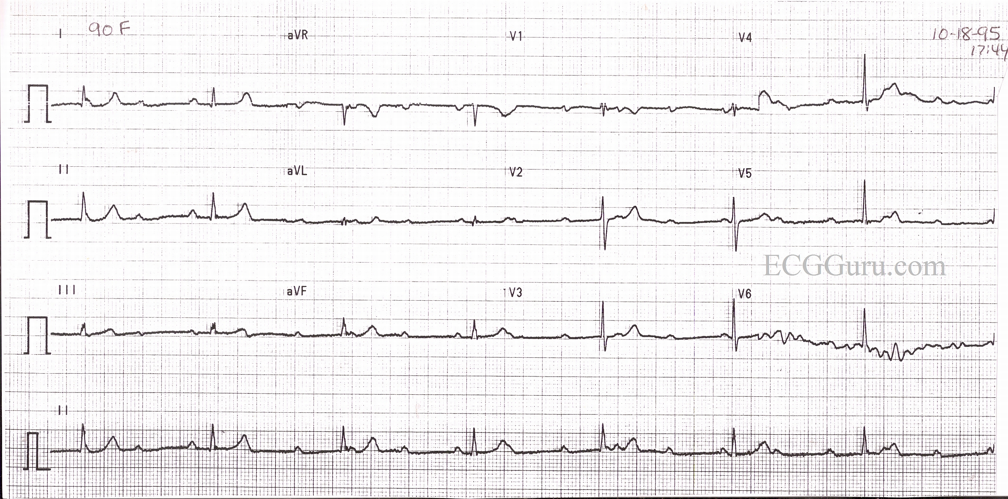
This ECG is from a 90-yr-old woman who complained of weakness and difficulty walking. The ECG shows an underlying sinus rhythm at 110 bpm. There is also a junctional rhythm at a rate of around 40 bpm. The QRS complexes are narrow, and there are no retrograde P waves. The two rhythms are unrelated, so this is complete heart block, or third-degree AV block with junctional escape.
There are several ways to prove conduction has occurred in AV block. One is for all the PR intervals to be the same (second-degree, Type II). Another is to see progressively-lengthening PR intervals in CONSECUTIVE P waves until one QRS is dropped (second-degree, Type I). Another time we can infer some conduction is when the QRS complexes are IRREGULAR, and not all P waves are conducted. This would imply a high-grade AV block where some beats are conducted and some are not. Complete AV block is diagnosed when there is an ESCAPE RHYTHM, either junctional or ventricular. Junctional escape rhythm would be regular, at a rate usually between 40 and 60, with narrow QRS complexes (assuming an absence of bundle branch block). Ventricular escape rhythm is regular, at a rate below 40, with wide QRS complexes.
The treatment for complete AV block is a permanent pacemaker.
Rate this content:

All our content is FREE & COPYRIGHT FREE for non-commercial use
Please be courteous and leave any watermark or author attribution on content you reproduce.
Comments
Good ECG
This is a good ECG for review. This is a rhythm that is not seen very frequently its nice to see it and get some history behind it.
Fred Vaccaro
AV Block does not Appear to be secondary to an MI
Since there is an absence of pathological Q waves can we assume that this block was the result of disease process and not an arterial occlusion?