Displaying 21 - 30 of 67
Dawn's picture

Inferior Wall M.I. With Right Ventricular M.I.

This ECG was recorded from a 75-year-old man with substernal chest pain and diaphoresis.  It shows a pretty classic picture of acute inferior wall M.I. The second ECG is a repeat tracing with the V4 wire moved to the V4 Right position, and it is positive for right ventricular M.I.  The patient was found to have a 100% occlusion of the right coronary artery, which was opened and stented in the cath lab.

There are several other examples of IWMI with RVMI in our archives, so we will confine this commentary to the ECG signs that make these tracings so typical of right coronary artery occlusion. Once you are familiar with the typical pattern of IWMI / RVMI, it is easy to see, even when the ST elevation is subtle (as this one certainly is NOT).

Signs of IWMI in these ECGs are

·         ST elevation in inferior leads II, III and aVF.

·         Reciprocal ST depression in leads I and aVL. 

Signs of RVMI in these ECGs are:

·         ST elevation in V4 right.

·         ST elevation in V1 without ST elevation in V2.

Dawn's picture

Simultaneous Occlusions in LAD and Diagonal

This ECG was obtained from a 35-year-old man who was complaining of crushing substernal chest pain which radiated down his left arm for the last ten minutes. He was diaphoretic, and described his pain as a “10” on the 1-10 scale. He got only modest relief from IV fentanyl.

He was transported to a full-service cardiac hospital, where he underwent angioplasty of simultaneous 100% occlusions of his proximal left anterior descending artery and diagonal artery. He was noted to have apical akinesia with a 35% ejection fraction.

He continued to improve following angioplasty, and was discharged home with an external defibrillator vest.

The ECG shows ST elevation in V2, V4, V5, and V6, which makes us suspect that the V2 and V3 wires were switched accidentally.  This reflects damage in the anterior wall of the LV. There is also very marked ST elevation in I and aVL, reflecting damage in the high lateral wall. There is reciprocal ST depression in the inferior leads aVF and III.  Fortunately, there are no pathological Q waves, which would indicate permanent damage from necrosis of the myocardium.

You can see films from his procedure in Heart Art, labeled “Simultaneous Occlusive Lesions in LAD and Diagonal”.

Dawn's picture

Inferior Wall M.I. With Wide QRS and Complete AV Block

This ECG is from a 66-year-old woman who called 911 for a complaint of chest pain for the past four hours. She also complained of nausea, vomiting, and diarrhea for that time. She was pale and diaphoretic, and her BP was 77/43 sitting up, improving to 90/54 reclining. She denied “cardiac” history.  Her medications included:  aspirin, an SSRI, cilostazol, amlodipine, umeclidinium and vilanterol inhaler, atorvastatin, levothyroid, and metoprolol. We don’t have a previous ECG.  The EMS crew followed their chest pain protocol and delivered the patient to a facility with an interventional cath lab, but they did not designate a “STEMI Alert” because of the wide QRS.  It is their protocol to use the term “STEMI Alert” only when no M.I. mimics, such as left bundle branch block, are present. 

What does this ECG show?     There is an underlying sinus rhythm at 75 bpm.  There is AV dissociation, with regular, wide QRS complexes at a rate of 44 bpm.   This meets the criteria for complete heart block (third-degree AV block).  The morphology of the QRS complexes meets the criteria for left bundle branch block (wide, upright in Leads I and V6, negative in V1).  At a rate of 44 bpm, several options for this escape rhythm are possible:  1)  junctional escape with LBBB, 2) junctional escape with intraventricular conduction delay due to AMI,  and 3) idioventricular escape rhythm.   Also, in the presence of IWMI, AV node ischemia is very likely, resulting in AV blocks at the level of the AV node.  CHB at the AV node would result in junctional escape rhythm, and CHB below that, in the fascicles of the bundle branches, would result in idioventricular escape. The issue for this patient, and ANY patient, is cardiac output, and we see several reasons for cardiac output to be lower:

·         Wide QRS

Dawn's picture

Brugada Pattern

This patient is a 50-year-old man with a history of epilepsy and early dementia. He had a VP shunt placed in the hospital and was then discharged home.  He became extremely weak, which was not characteristic of him, and 911 was called.  He was transported to the hospital uneventfully. He was found to be afebrile.

This ECG shows a “classic” Brugada pattern. Brugada Syndrome is a hereditary disease that is associated with a high risk of sudden cardiac death. It is due to a mutation in the sodium channel gene (channelopathy). The ECG characteristics are:

·         ST elevation in V1 through V3 of at least 2 mm at the J point in the right precordial leads (V1-V3).

·         Coved upward ST segments with  negative T waves in the affected leads.

Brugada Syndrome is diagnosed when the ECG pattern exists with one of the following:

·             Polymorphic ventricular tachycardia (VT).

·             Documented ventricular fibrillation.

Dawn's picture

Anterior Wall M.I. With Bifascicular Block

This ECG is taken from an 82-year-old man who called 911 because of chest pain.  He has an unspecified “cardiac” history, but we do not know the specifics. 

WHAT IS THE RHYTHM?  The heart rate is 69 bpm, and there are P waves before every QRS complex. The underlying rhythm is regular, with one premature beat that is wide without a P wave.  The PR interval is slightly prolonged at .25 seconds.  The rhythm is normal sinus rhythm with first-degree AV block and one PVC. 

WHY THE WIDE QRS?   The QRS complex is wide at .14 seconds. The QRS in V 1 has a wide R wave after a small Q wave.  This in consistent with right bundle branch block pattern, with loss of the normal initial small r wave (pathological Q waves).  The diagnosis of RBBB is further corroborated by the wide little S waves in Leads I and V6.  The QRS frontal plane axis is -66 degrees per the machine, and clearly “abnormal left” because the QRS in Lead II is negative, while the QRS in Leads I and aVL are positive.  This is left anterior fascicular block, also called left anterior hemiblock.  The combination of RBBB and LAFB is a common one, as the two branches have the same blood supply.  It is also called bi-fascicular block. 

WHAT ABOUT THE ST SEGMENTS?  The ST segments in leads V2 through V6 are elevated, and their shape is very straight, as opposed to the normal shape of coved upward (smile). Even though the amount of ST elevation at the J points appears subtle, the shape of the segments, the fact that they appear in related leads, and the fact that the patient is an elderly male with chest pain all point to the diagnosis of ANTERIOR WALL ST elevation M.I. (STEMI).  Additional ST changes include a straight shape in Leads I and aVL and ST depression in V1 and aVR.  

PATIENT OUTCOME  The patient was transported to a cardiac center, where he received angioplasty in the cath lab.  The left coronary artery was found to be occluded, and was repaired and stented.  He recovered without complications and was sent home in a few days.

Dawn's picture

Incorrect Machine Interpretation

This ECG is presented as an example of INCORRECT MACHINE INTERPRETATION.  While there are many abnormalities in this ECG, it does not represent a paced rhythm. While there are exceptions, most paced rhythms represent either AV sequential pacing, right ventricular pacing, or bi-ventricular pacing.

RECOGNITION OF A PACED RHYTHM

Recognizing a paced rhythm can be difficult in some cases. Because pacemakers now have so many programmable features, there is a wide variety of ECG changes associated with them.  Pacer “spikes” can be difficult to see in all leads.  Finding evidence of the device on the patient’s chest or via patient history is a big help in reminding us to scrutinize the ECG for paced rhythm.

An AV sequential pacemaker or a right ventricular pacemaker will pace the ventricles via the right ventricle.  This produces a WIDE QRS and a leftward axis, often causing Leads II, III, and aVF to be negative and aVL and aVR to be positive.  Along with the wide QRS, we will see DISCORDANT ST CHANGES.  That is, there will be ST depression and T wave inversion in leads with positive QRS complexes and ST elevation and upright T waves in leads with negative QRS complexes.

Bi-ventricular pacing can be a little more complicated to recognize, as the QRS can be narrow, with signs of fusion between the wave produced by the LV electrode and the RV electrode.

The frontal plane axis is usually far right – aVR will be positive.  Lead I will be negative.

The machine is wrong:  there is no indication of a pacemaker, and P waves are present, even though they are not noted in the "PR Interval" or "P Axis".

SO, THIS IS NOT A PACED RHYTHM – WHAT IS IT?

Dawn's picture

Acute M.I. In Patient With Pacemaker

This ECG is taken from an elderly man who has a history of complete heart block and AV sequential pacemaker.  On the day of this ECG, he presented to the Emergency Department with chest pain and shortness of breath. His vital signs were stable and within normal limits.  We do not have information about his treatment or outcome. 

I don’t see spikes.  How do we know this is a paced rhythm?  The ECG clearly shows the presence of an AV pacemaker.  There are very tiny pacer “spikes”, probably best seen in Leads III, aVF, aVL, and most of the precordial leads.  Other ECG signs that this is a paced rhythm are:  wide QRS at about .16 seconds (160 ms); abnormal left frontal plane axis; regular rhythm with AV dissociation (there are P waves seen occasionally that have no fixed relationship to the QRS complexes).  Also, V6 is negative.  That rules out left bundle branch block unless the electrodes are misplaced.  There are no capture beats in this strip.  The patient appears to be, at least right now, 100% dependent on the paced rhythm. 

Why does the presence of a pacemaker make it harder to diagnose an M.I. from the ECG?  Wide-QRS rhythms, such as right-ventricular paced rhythms, left bundle branch block, and ventricular ectopic rhythms, usually have “discordant ST and T wave changes”.  That is, when the QRS is positive (upright), the ST and T wave are negative.  The reverse is also true:  when the QRS is negative and wide, the ST and T wave changes are positive (ST elevation).  This is not true for right bundle branch block because the conduction delay that causes the widening of the QRS is in the right ventricle, and the ST segment is reflecting the LEFT ventricle’s repolarization.  Discordant ST changes can make it difficult to determine from the ECG alone that there is an ST elevation M.I. (STEMI).  Diagnosis usually must be made from patient presentation, ECG changes over time, and cardiac enzymes – or more definitively from cardiac angiogram. Pacemakers that produce narrow QRS complexes do not cause discordant ST changes. 

Dawn's picture

Inferior-lateral M.I. With QRS Fragmentation

SUBTLE ST CHANGES   This ECG was obtained from an 87-year-old man who was experiencing chest pain.  Due to the subtle ST elevation in Leads II, III, aVF, V5, and V6, (inferior- lateral walls) the ECG was transmitted to the hospital by the EMS crew, and the cath lab was activated.  The patient denied previous cardiac history. 

In addition to the subtle ST elevation, there is ST depression in V1 through V4, which represents a reciprocal view of the injury in the inferior-posterior-lateral wall.  Because the anterior wall is superior in its position in the chest, it is opposite the inferior/posterior wall, and can show ST depression when the inferior-posterior area has ST elevation. This ECG was the 6th one done during this EMS call.  Prior to this one, the ST segments were elevated less than 1 mm.  This is a good example of the value of repeat ECGs during an acute event.  

RIGHT VENTRICULAR M.I.?     This ECG was done with V4 placed on the right side, to check for right ventricular M.I., which is a protocol for this EMS agency. When the right coronary artery is the culprit artery (about 80% of IWMIs), RVMI is likely.  In RVMI, we would usually see reciprocal ST depression in Leads I and aVL, but the STE is very subtle here, so the depression would likely be also.  When the culprit artery is the left circumflex artery (<20%), lateral lead ST elevation is more likely, as we see here in V5 and V6. 

WHAT ABOUT RHYTHM?     The rhythm is sinus with PACs.  PACs are considered to be benign in most situations, but in a patient with acute M.I., any dysrhythmia can be concerning. The QT interval, measured as QTc (corrected to a heart rate of 60 bpm), is slightly prolonged at .458 seconds (458 ms).  Over .440 seconds is considered prolonged in men, and over .500 sec. places the patient at increased risk of developing torsades de pointes.  CAD and myocardial ischemia can lead to this modest increase in QTc.

Dawn's picture

Teaching Series: Acute Anterior Wall M.I.

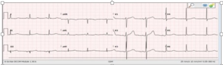
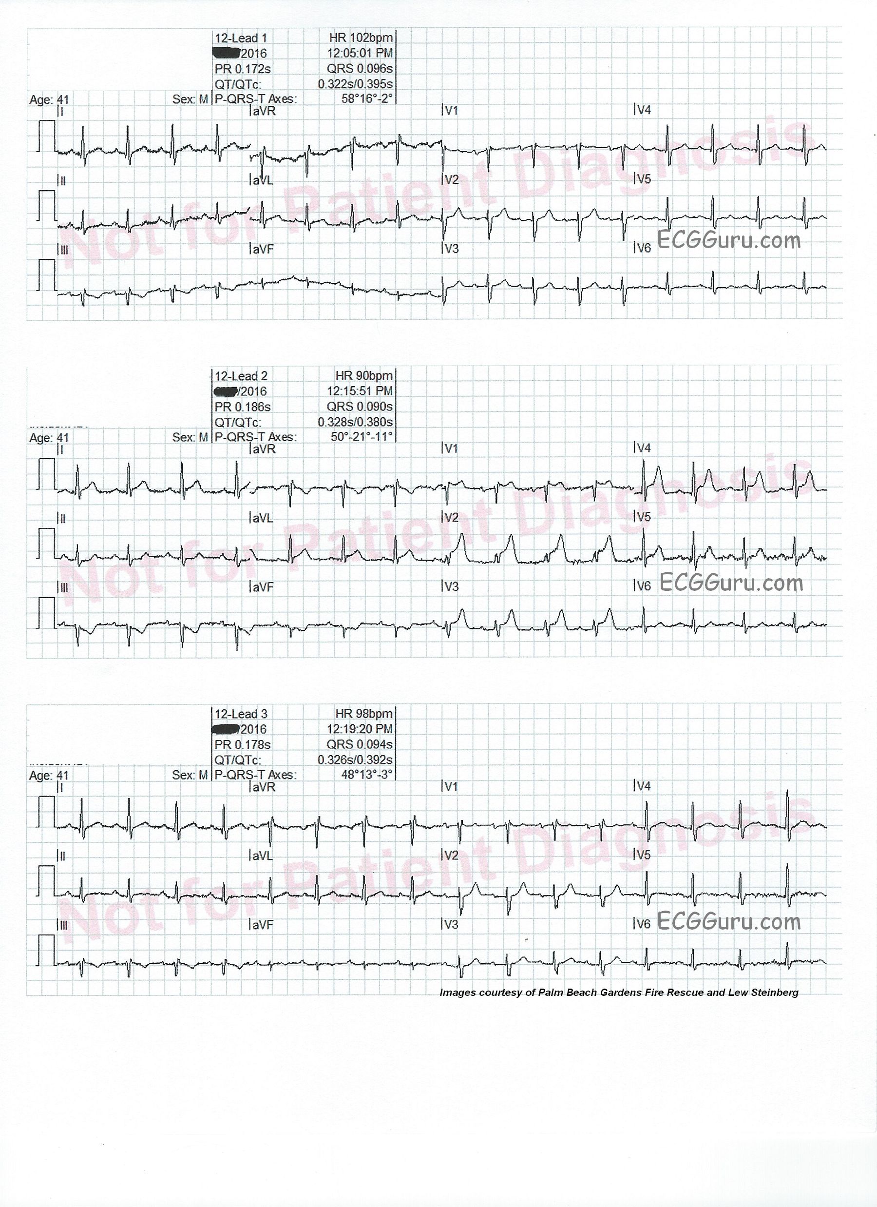
Intermittent chest pain.     This series of three ECG were taken from a 41-year-old man with a two-week history of intermittent chest pain.  At the time of the first ECG, 12:05 pm, he was pain-free.  We see a sinus tachycardia at 102 bpm, and has just come under the care of paramedics. There is a very subtle ST sagging and T wave inversion in Lead III, and no other ST changes. He had an uneventful trip to the hospital.

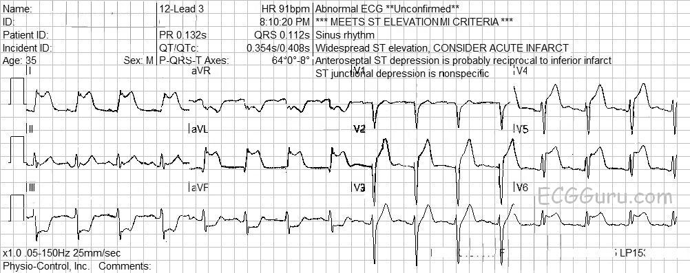
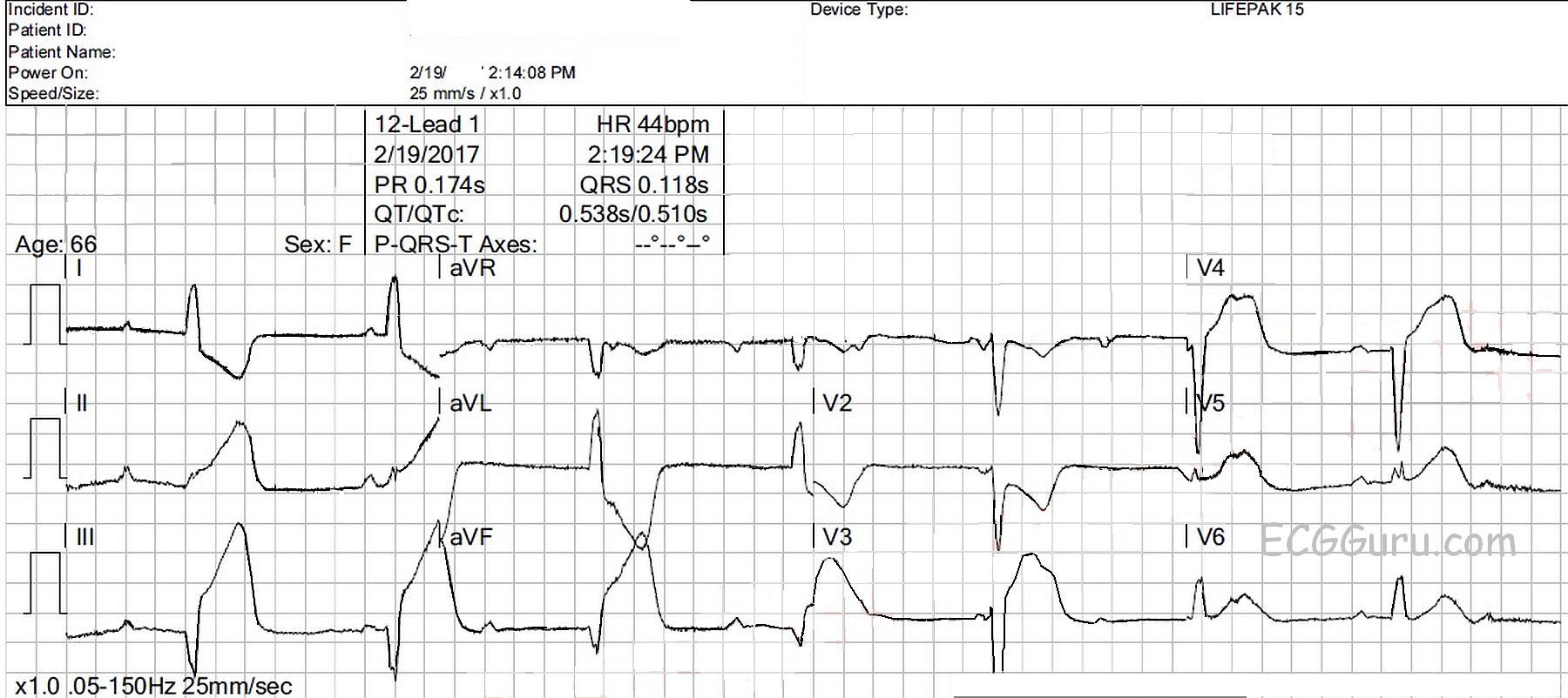
On arrival at the Emergency Department, just before he was unloaded from the ambulance (12:15), he experienced chest pain.  An ECG was obtained, which shows ST elevation in V1 through V4, as well as in Leads I and avL.  There are reciprocal ST depressions in Leads III and aVF. 

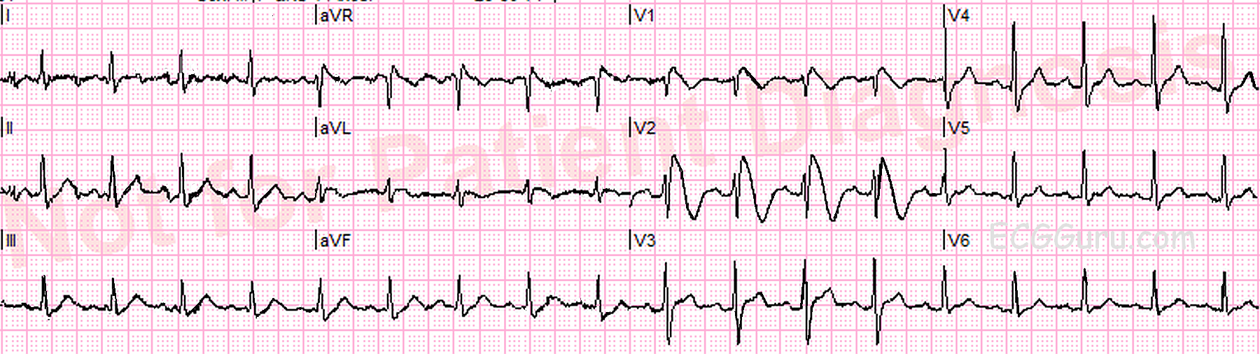
The patient was taken into the ED, where his symptoms abated, and a third ECG was obtained (12:19). The third ECG looks very much like the first one. V5 and V6 have T waves that appear flat, or even inverted, but there is some baseline artifact making it hard to see them. 

Diagnosis confirmed       Based on the patient’s presentation, and the second ECG, he was taken immediately to the cath lab. A 100% occluding lesion with a clot was discovered in the mid LAD.  The clot was removed with suction, and the lesion stented.  A 40% narrowing was discovered in the RCA.  

Repeat ECGs whenever possible       This series of ECGs offers a compelling argument for performing repeat ECGs.  This can be especially important when symptoms are waxing and waning.  Sometimes, a clot can completely occlude an artery, then “float” to another position, allowing blood flow to resume.  Sometimes, the artery constricts around the lesion, causing occlusion, then relaxes.  Had the rescue crew not repeated the ECG at 12:19, there may have been a delay in this young patient receiving interventional care.

Dawn's picture

Subtle ST Changes

This ECG is from a 65-year-old woman who presented to the Emergency Department with a complaint of chest pain.  We have no other clinical information. 

There are several subtle, but real, abnormalities on the ECG that should be evaluated in conjuction with her clinical situation.  This is a good ECG to discuss with your more advanced students who have mastered recognition of blatant ST elevation M.I. (STEMI).  But, let’s start with what is NORMAL here. 

NORMAL FEATURES        The normal findings are:  normal sinus rhythm at about 68 bpm.  The rhythm is regular without ectopy. The intervals are within normal limits.  The frontal plane axis is normal. The T waves are all upright. 

ABNORMAL FEATURES         There is subtle ST segment elevation in Leads V1 through V3, and in I and aVL.  The shape of the ST segments is concave upward, or normal.  In a young, asymptomatic patient, we probably would not be at all concerned about this amount of ST elevation.  However, this is an older patient with chest pain. In the chest leads, the R wave progression is interrupted, as the QRS goes abruptly from negative to positive in Lead V3.  This could be due to loss of r waves ( pathological Q waves forming) in V1 and V2 lead placement, or something else. 

Pages

All our content is FREE & COPYRIGHT FREE for non-commercial use

Please be courteous and leave any watermark or author attribution on content you reproduce.