During our summer break, we are reprising a few of the best ECGs from our archives, to give you a chance to comment or to ask questions.
This ECG was taken from a 52 year old man who was complaining of chest pain, with a history of severe multi-vessel disease. He has a history of M.I. and states he has five coronary stents.
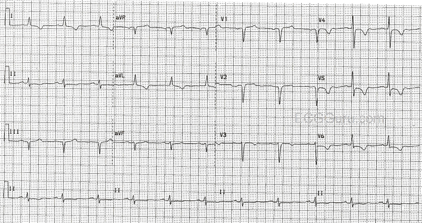
His pain was partially relieved by Ntg., and he was given aspirin in the field, and then IV Ntg., Integrelin, and morphine before being sent to the cath lab. This ECG shows T wave inversions with coved upward ST segments, but no ST elevation in the lateral leads: I and aVL, and the anterior-lateral leads, V3 through V6. This represents the territory covered by the left coronary artery, and points to a lesion in the proximal portion of the artery. Also in this ECG are pathological Q waves in right side leads, III, V1 and V2.
In the cath lab, he was discovered to have a ruptured plaque in the proximal LAD, with some blood getting through a very narrow channel. He was referred for coronary artery bypass surgery the next day.