
Submitted by Dawn on Wed, 05/22/2013 - 20:11
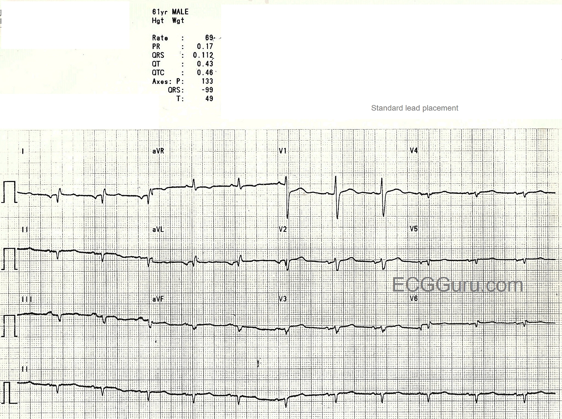
This middle-aged man presents to the Emergency Dept. complaining of shortness of breath and fever. Blood work, chest xray, and ECG are ordered. When the ER Tech obtains the 12-Lead ECG, he is puzzled by the following findings:
Low voltage in the chest leads V2 - V6
Abnormal right axis of P waves with negative P waves in Leads I and aVL
Abnormal right QRS axis with an upright aVR.
Seeing his puzzled look, the patient says, "Oh, I forgot to tell you, I have mirror-image dextrocardia!"
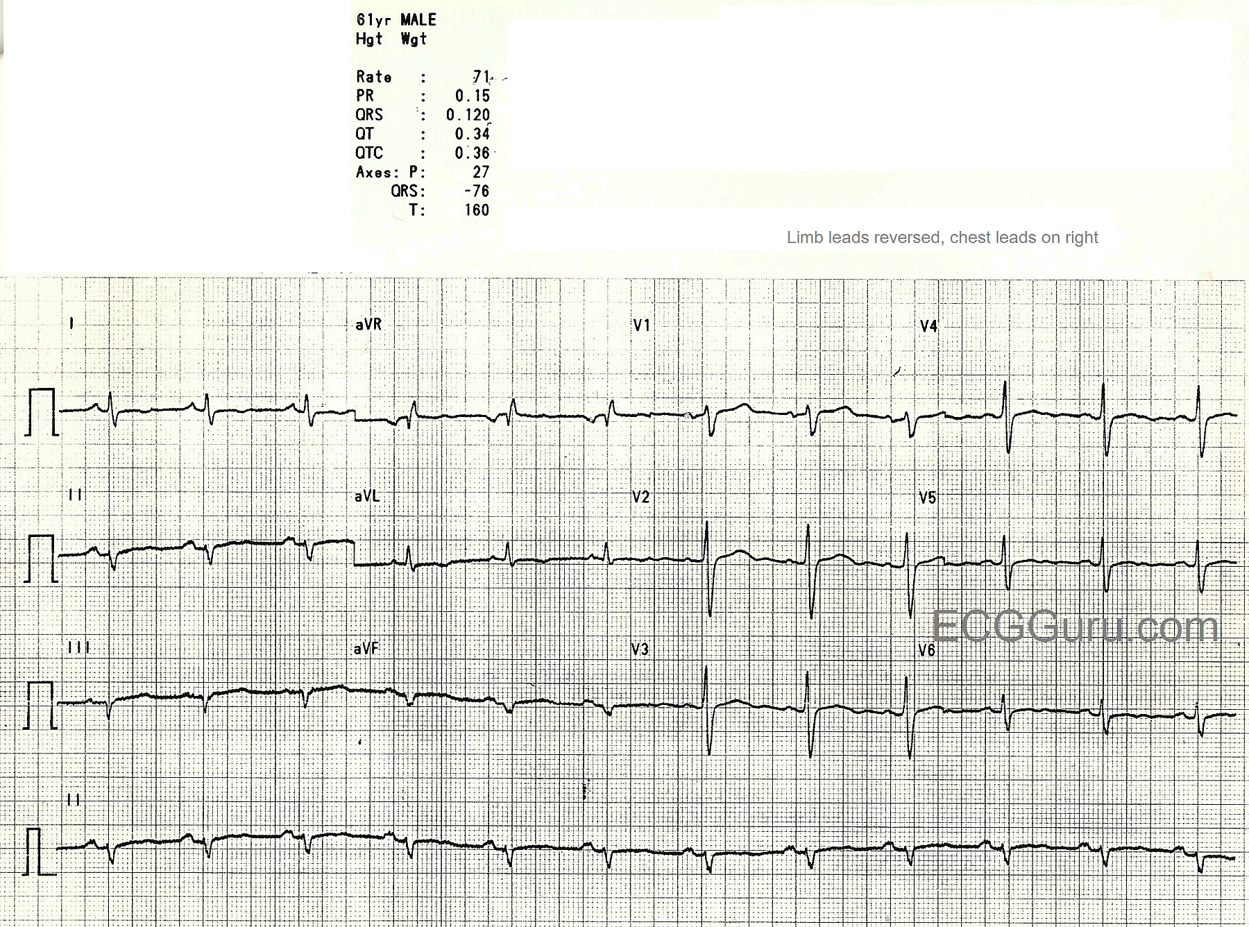
After consulting with the ED physician, the Tech reverses the arm leads and the chest leads and obtains the second ECG.
The P wave axis is now correct, although there is still derangement of the QRS axis. The QRS complexes in the chest leads look more normal, but do not show the usual R wave progression of the normal ECG.
In dextrocardia, the heart is located on the right side of the chest. Approximately 1 in 12,000 people have dextrocardia. There are several versions of the anomaly.
MIRROR-IMAGE DEXTROCARDIA: The heart is a mirror image of normal, with the atrial and ventricles reversed. 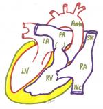
The placement of the great vessels is usually normal. Often, the visceral organs are also reversed (situs inversus). 
On xray, the gastric gas bubble can be seen on the patient's right. The heart usually functions normally in this situation. You should do a "reverse" ECG, with limb leads and chest leads reversed.
MIRROR-IMAGE DEXTROCARDIA: The heart is a mirror image of normal, with the atrial and ventricles reversed. The placement of the great vessels is usually normal. Often, the visceral organs are also reversed (situs inversus). On xray, the gastric gas bubble can be seen on the patient's right. The heart usually functions normally in this situation. You should do a "reverse" ECG, with limb leads and chest leads reversed.
DEXTROVERSION (Isolated Dextrocardia): The heart is on the right, but the atria and ventricles are arranged normally. This is often associated with other heart malformations. Some patients with dextrocardia will have other congenital anomalies and syndromes.
Often, however, the heart and other organs will be perfectly normal, just reversed.
TIPS: If you think you are seeing dextrocardia on the ECG, but the patient has no history of it, first check your arm leads for reversal. The chest xray offers a valuable insight also.
When defibrillating or pacing the patient with dextrocardia, place the pads opposite of normal (apex pad over right lower chest).
For more about dextrocardia, check out these links: Life in the Fast Lane, Medline Plus.
Related Terms:
Rate this content:
All our content is FREE & COPYRIGHT FREE for non-commercial use
Please be courteous and leave any watermark or author attribution on content you reproduce.