Dawn's picture

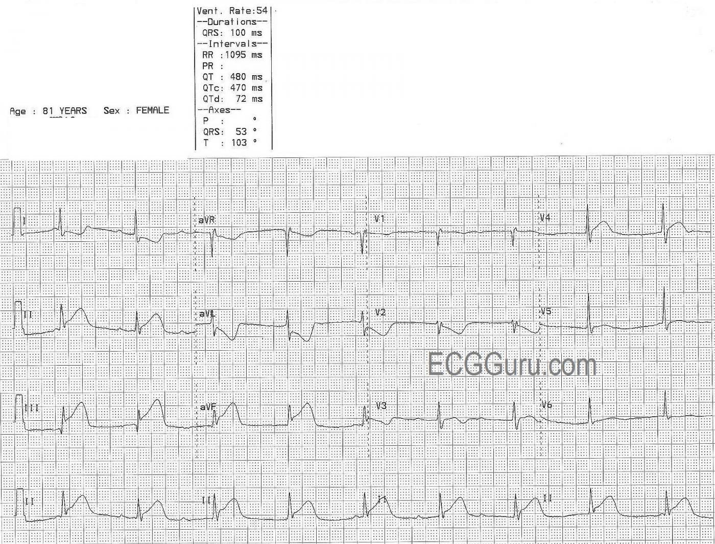
This ECG is from an 81 year old woman with an extensive history of coronary artery disease.  She was experiencing chest pain at the time of the ECG.  We can clearly see ST elevation in Leads II, III, and aVF, indicating an inferior wall ST-elevation M.I. (STEMI).  There are reciprocal ST depressions in Leads I and aVL.  There are subtle and less specific ST changes in V1 (flat ST and T), V2 (ST depression), V3 (ST elevation and inverted T wave), and V4 through V6 (slight ST elevation).  The flat, horizontal shape of most of the ST segments is another clue to her CAD.  What coronary artery do you think is the culprit for the ST elevation?

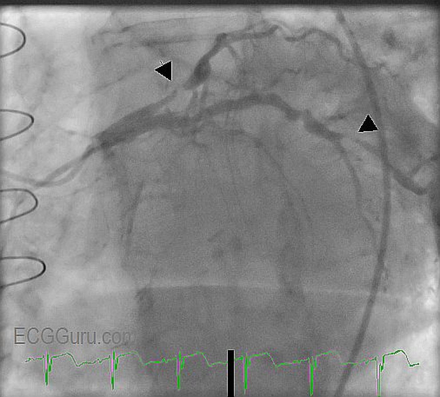
This patient received coronary angiography, so we do not have to guess at where her lesions are.  She was found to have an occluded left internal mammary artery (LIMA) graft.

The left anterior descending coronary artery (top of view) is very diseased, with some extremely narrow areas.  The circumflex artery is large and covers a lot of area, but it, too, is very diseased, with a critical occlusion (marked with arrow).  On this day, the circumflex artery received angioplasty and stents.  The patient's right coronary artery was patent.

In approximately 85% of the population, the RCA supplies the inferior wall of the heart.  In most of the rest, a branch of the circumflex supplies the posterior/inferior wall.

This patient also has a very interesting arrhythmia.  We see P waves (numbered) that appear regular for three beats, then we see no P waves for a pause.  Then, three more P waves appear.  There is no readily-seen "hidden" P wave in the ST segments or T waves.  Even though there appear to be "progressively prolonging PR intervals", the PR intervals of each group of three do not match the other group of three.  The first "PRI" - P wave number 1- and the last one - P wave number 7 - appear too short to be normal PR intervals.  The ventricular rate is regular, and the QRS complexes are slightly wide at  .10 sec.  The rate is 54 bpm.  This suggests junctional rhythm.

We are eager to hear your comments regarding this rhythm.

Rate this content: 
Average: 3.9 (7 votes)

Comments

ekgpress@mac.com's picture

     VERY interesting case by Dawn - both for the rhythm as well as the 12-lead ECG. It is always insightful when we have "the answer" (= cath films) - since we can then KNOW where the lesion is. As always - I do my best NOT to bias my interpretations - so I had no idea of what the cath showed until AFTER I interpreted the tracings. One learns more this way.

I will first address the 12-LEAD ECG - leaving the rhythm for later. MY IMPRESSION (prior to looking at cath film results):
  • Supraventricular rhythm (since the QRS is narrow) - which is junctional. You CAN interpret ST-T wave changes with junctional rhythm.
  • Acute inferior MI (marked ST elevation in II, III, aVF ). Given that ST elevation is by far greatest in III > II - and that ST depression is clearly more in aVL > I - this would suggest proximal RCA (Right Coronary Artery) occlusion IF this was a new event in a patient without prior disease.
  • There is an IRBBB (Incomplete Right Bundle Branch Block) pattern - in that the QRS is not wide; there is a Qr in V1 and rSr' in V2 with narrow terminal s in I, V6.
  • Suspect acute posterior involvement from the complete mirror image picture in V2 of the inferior ST elevation.
  • The relatively flat ST segment in V1 would make me think of associated RV involvement IF this was a 1st event in a patient without CAD.
  • BUT - there is ST coving with T inversion in V3, and definite ST elevation in V4 - which would NOT be expected if this was a new 1st event due to proximal RCA occlusion .... So there is a bit of a "disconnect" in terms of which coronary artery is likely to be occluded ....
  • Clinically - this is not a problem - since it is obvious that there is a new acute stemi = Take the patient to cath!
  • KNOWING that this patient has a history of extensive coronary disease WITH prior bypass surgery resolves things. This is not a native coronary anatomy. The patient apparently has a dominant left circumflex with diffuse disease in that vessel and in her LAD. Bypass surgery, collateralization, prior events all explain the "disconnect" in terms of involved areas that I postulated earlier.
  • MORAL of STORY: When the 12-lead suggests conflicting information as to which coronary artery might be occluded with an acute event - the reason may simply be that there is extensive coronary disease with prior revascularization and collateralization. 
THE RHYTHM:
  • Knowing this patient is having an acute STEMI helps narrow down the likely possibilities for this supraventricular rhythm.
  • To me - the BIGGEST CLUE to the rhythm is that the ventricular rhythm is precisely regular.
  • HINTS: It is best to DOWNLOAD this tracing and to print it out. Use Calipers. The ECG is larger when you download the image and print it out. NUMBER each QRS complex in the long lead II rhythm strip at the bottom.
  • As Dawn mentions - a number of P waves are seen. Others are not seen. IF I set my calipers to an interval equal to the P-P for 2 P waves that ARE seen - I can pretty much walk the calipers through the entire tracing. I agree that there is no notching with ST-T waves following the 3rd and 4th QRS complexes - nor after the 7th or 8th QRS complexes - and I DO have to slightly change my caliper length in order to get this to walk out. This to me suggests probably sinus arrhythmia. 
  • The fact that the QRS remains narrow and precisely regular throughout with places where I KNOW the P wave is NOT conducting (the PR is too short to conduct preceding the 1st and 9th QRS complexes - and probably also before the 5th QRS) - means the underlying junctional rhythm persists throughout. 
  • PEARL: When there is 2nd degree AV block with only intermittent AV dissociation - there should be a CHANGE in the R-R interval. Usually the slightly shorter R-R signals which QRS is conducting. But the R-R stays constant throughout this rhythm strip! Therefore - this is complete (3rd degree) AV block. The atrial rate is ~ 65/minute (with slight variability) and the junctional rate an appropriate 55/minute.
  • The KEY to diagnosing 3rd degree AV block is that there is complete AV dissociation with NO P wave conducting despite having adequate opportunity to conduct. The ventricular rate needs to be slow enough (usually <50/minute) for P waves to occur in all phases of the R-R cycle in order for us to be sure a rhythm is 3rd degree. The reason this rhythm strip is so challenging - is that the ventricular rate isn't quite that slow .... so it is difficult to be certain P waves (especially when you can't see them all ...) have add "adequate opportunity" to conduct. So while technically I'd like to see a bit more rhythm monitoring to be absolutely certain there is no conduction - I think the 9 beats of absolutely regular R-R interval seen here is strongly suggestive that this indeed is complete AV block.
NOTE: For those wanting a primer on "Which Vessel" is likely to be occluded with acute STEMI - Please CLICK HERE.
 

Ken Grauer, MD  www.kg-ekgpress.com   [email protected] 

All our content is FREE & COPYRIGHT FREE for non-commercial use

Please be courteous and leave any watermark or author attribution on content you reproduce.