Dawn's picture

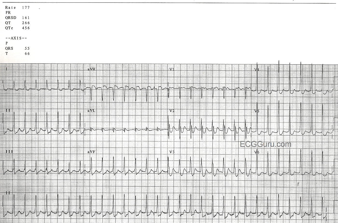
This ECG is from a man in his 60's who is experiencing chest discomfort and palpitations.  The onset of the rapid heart rate and the symptoms was sudden, while he was at rest.  The rate did not slow when he was placed on oxygen, given IV fluids, and rested further. The rate is 177 / min.  

The rhythm is AV nodal reentry tachycardia (AVNRT), which is one of the rhythms that falls into the category of paroxysmal supraventricular tachycardia (PSVT).We can see signs of retrograde P waves in some leads (II, III, aVF, V1).  AVNRT is caused by a reentry circuit in the AV node.


Some instructors teach students that sinus tach is approximately 100-150 per minute, and atrial tach is usually 150-250 per minute.  If students only learn about differentiating these two rhythms by the rate difference, it will cause later problems.  Of course, there is actually an overlap in rates between the two rhythms.  For example, a febrile, dehydrated infant could easily reach this rate and be in sinus rhythm.  A young, healthy person on a treadmill could, too.  Clues to the ectopic origin of this rhythm are:  sudden onset (unfortunately, not witnessed here), regular rhythm with unwaivering rate, and the patient's situation (symptoms while at rest, no obvious reason for sinus tach).  Of course, we need to teach to the level of our students' abilities.  Consider whether they just memorizing rhythms criteria now, or are they learning about re-entry?

There are many different mechanisms of supraventricular tachycardia, and they differ on an electrophysiologic basis.  Depending upon the level of your students, you will have to decide how much detail to teach.  There are many resources on this site, including the ECG Archives, Ask the Expert, Jason's Blog, and Favorite Sites that will help you if you want to go into more detail.  For many health care providers, the entire category of "PSVT" is treated the same initially, and there is no need to differentiate the types of PSVT.


Rate this content: 
Average: 3.8 (5 votes)

Comments

ekgpress@mac.com's picture

 
As per Dawn  the 12-lead ECG shown is of a regular SVT (SupraVentricuar Tachycardia) rhythm at a rate of ~180/minute without clear evidence of sinus P waves. I’ll add a few comments to Dawn’s discussion:
  • The principal Differential Diagnosis of a regular SVT without sinus P waves includes 3 entities: i) Sinus tachycardia (in which sinus P waves may be hiding within the preceding T wave); ii) Atrial Flutter; and iii) AVNRT ( = AV Nodal Reentrant Tachycardia  formerly known as PSVT = Paroxysmal SupraVentricular Tachycardia). Of course  there are other entities to include in the differential  but they are far less common. In a nonreferral ambulatory and/or hospital population  in my experience, >90% of all regular SVT rhythms at a rate >140/minute will be one of the above 3 entities …
  • The heart rate (about 180/minute should provide a BIG clue as to etiology. A rate over 170/minute makes sinus tachycardia far less likely (especially in an older adult). Please note that I did NOT say “impossible”  but far less likely. It IS possible to have faster rates for sinus tachycardia  but common things are common  and a rate >170/minute starts me thinking down the path of a reentrant tachycardia (especially if onset is sudden). 
  • The heart rate of ~180/minute also makes AFlutter far less likely  since the most common AV response to untreated AFlutter is 2:1  and the atrial rate of flutter in adults usually does not exceed 350/minute (most often closer to ~300/minute). So the rate of 180/minute seen here make AFlutter that much less likely.
  • I can become even more comfortable with the diagnosis of AVNRT for this case  because I agree with Dawn that we DO see probable retrograde P waves (notching the terminal end of the QRS in each of the inferior leads  and, notching the beginning of the ST segment in leads aVR and V1). Thus, the RP’ interval ( = distance from the QRS until the point where the retrograde P wave is seen) is SHORT  due to likely retrograde conduction back to the atria over the “fast” pathway  which is the most common occurrence with AVNRT. In contrast  we do NOT see any suggestion of flutter waves (I set my calipers to precisely HALF the R-R interval  and look to see if anything can be marched out, and it cannot … ).
  • While Dawn’s comment that many regular SVT rhythms are treated the same initially is true  KNOWING (or being almost certain, as I am here) that the mechanism of this rhythm is AVNRT is indeed helpful to me  because it tells me how far to persist down a given course of treatment. Adenosine usually converts this rhythm. But Adenosine won’t usually convert AFlutter  and of course, it won’t treat Sinus Tachycardia. So I have “more ammunition” to support an approach of perhaps repeating Adenosine if it is not effective with the initial dose. Adenosine doesn’t work 100% of the time  so knowing this is almost certainly AVNRT might lead me to trying another AV nodal blocking agent if Adenosine didn’t work. This is not to say that we wouldn’t do the same empirically if we didn’t know the mechanism  but it IS to say that I’d have more confidence doing so knowing the almost certain mechanism of this rhythm.
  • Finally  there is a LOT of ST depression on this tracing! In addition - the ST segment is elevated in lead aVR. While this combination of ST-T wave findings often suggests severe (even left main) disease  you cannot rely on that dictum given the marked tachycardia  since AVNRT is commonly associated with similar amounts of diffuse ST depression that is NOT due to coronary disease, and which resolves after conversion of the rhythm to sinus. Therefore  REPEAT the ECG after sinus rhythm is restored.
  • Be sure to look at the terminal portion of the QRS after you repeat the ECG! That notching I mentioned above should be gone IF it in fact truly was the result of retrograde atrial activity during AVNRT.
 ------------------------------------------------
For those wanting more on AVNRT  Please check out:
  • My ECG Blog #97 ( = ECG Video #3 GO TO  https://youtu.be/5E3_MKuvr9c - 
  • My 3-part ECG Video on Cardiac Arrhythmia. For a timed and linked Table of Contents to this hour-long presentation  GO TO   http://tinyurl.com/Rhythm-Mgmt-Contents - 

Ken Grauer, MD  www.kg-ekgpress.com   [email protected] 

All our content is FREE & COPYRIGHT FREE for non-commercial use

Please be courteous and leave any watermark or author attribution on content you reproduce.