Dawn's picture

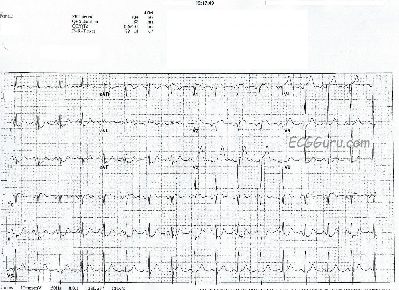
A 78-year-old woman complained of nausea and diaphoresis.  Paramedics in the field found that her 12-lead ECG showed ST elevation in V1 through V4, aVL, and aVR.  The patient denied chest pain and also denied any cardiac history.  She did not want to be transported to the hospital, but thankfully, the paramedics understood that this was not an option, and convinced her to go.

She was taken to a cardiac facility as a STEMI Alert, was evaluated in the cath lab, and sent immediately to the O.R. for coronary artery bypass surgery.  She had severe multi-vessle disease and a lesion in her proximal left coronary artery.  No other details of the cath results are known.

Some important teaching points:

  • there is subtle ST elevation in V1 and V2, but the SHAPE of the ST segment is suspect, with flattening and almost a coving upward shape in V1.  Normal ST segments are convex downward, like a smile.
  • there is nearly complete loss of r waves in V1 and V2, and V3 and V4 have very small r waves.  This signals impending pathological Q waves, a sign of necrosis of the myocardium.  Necrotic muscle does not contract.
  • there is slight ST segment elevation in aVR.  Along with STE in V1, this is a marker for proximal LCA or left main occlusion.
  • the ST elevations in V3 and V4 are more pronounced, and easily meet STEMI guidelines:  currently 1.5 mm of elevation in V3 and 1 mm of elevation in V4 for a woman.
  • there are reciprocal ST depressions in II, III, and aVF - common in AWMI.
  • aVL has slight STE, along with inverted T waves. Somewhat surprisingly, there is no ST depression in Lead I.  This indicates high lateral wall injury.
  • the patient has a "hint" of the criteria for LVH:  her S wave in V3 + her R wave in V5 = about 33 mm, and there is depression in V6.  A stretch to call it "LVH", but possibly a sign of left ventricular strain because of the acute M.I.
  • there are atrial abnormalities suggested by the tall, peaked P waves in Lead II, the "M" shaped P waves in Lead III, and the inverted P waves in V1 and V2.  Possibly bi-atrial dilation and stress brought on by the M.I.?  An echocardiogram would be a better test for this.
  • the heart rate, at about 90 bpm, reflects NSR but is a cause of more stress on an overworked, injured heart.

This is a great teaching ECG, and we hope the Gurus out there will add even more interesting points to consider.

Rate this content: 
Average: 4.6 (5 votes)

Comments

ekgpress@mac.com's picture

     I am glad there is cath confirmation for the coronary anatomy relevant to this tracing - as I otherwise was having difficulty explaining ECG findings by a single lesion. I'll expand on a number of points brought up by Dawn:
  • The history sounds consistent with an ongoing acute event - a 78-year old woman with nausea and diaphoresis, albeit denying chest pain. In this context - the ST segment elevations and depressions described are of definite concern.
  • Regarding terminology - there is a lack of consensus. The problem with use of the word "convexity" to describe ST segment shape is that one never knows IF the observer is describing the ST segment from above or below. Rather than "downward convexity" - I prefer the term, "coved" for ST segments in a "frowny" configuration (as seen in leads aVL and V1 of this tracing). The opposite shape is an upward concavity = "smiley" configuration - as illustrated by the ST segments in leads V3,V4 of this tracing. So normal ST segments are best described as "concave up" (rather than convex down).
  • Lack of consensus extends to terminology about wall location. Traditionally, lead aVL has been described as a "high lateral lead". Newer studies using MRI correlation instead suggest more of a mid anterior location for the area of the heart corresponding to ST deviation in lead aVL (Bayes de Luna - Rev Esp Cardiol 60:683-689, 2007). This makes sense when one considers results of a study by Birnbaum et al - which correlated likely location of the acutely occluded culprit artery when ST elevation was seen in lead aVL (as occurs in this tracing) - based on which other anterior leads showed either ST elevation or depression (Am Heart J 131:38-42, 1996).Thus, the finding in this case of subtle-but-real ST coving and elevation in lead aVL - in conjunction with more ST elevation in V1 > aVR, in conjunction with J-point ST elevation in V2-thru-V4 - is strongly suggestive of a proximal LAD lesion. For a SUMMARY of this useful Birnbaum reference - CLICK HERE
  • What puzzled me about this tracing - was the disproportionately large amount of ST depression in each of the inferior leads. So while there is J-point ST elevation in V1-thru-V4 - the shape of the ST segment is concave up (rather than coved) in V3,V4 - and the overall amount of J-point elevation is not nearly as much as I'd expect given how deep reciprocal ST depression in the inferior leads is. It was in fact this deep inferior ST depression in conjunction with non-"strain"-like ST depression in V5,V6 that made me think severe multi-vessel disease was likely in this 78-year old woman, especially given accompanying ST elevation in aVR and V1.
  • I would not call LVH on this tracing. As Dawn mentioned - R wave progression is delayed (no more than a tiny r wave up to lead V4). Whether this is truly the result of ongoing anterior infarction (null vector from loss of anterior forces) vs a lead placement issue is uncertain. Assuming delayed transition IS the result of anterior infarction - determination of LVH becomes problematic. The more anterior forces are lost (from anterior MI) - the deeper anterior S waves are likely to be independent of any increase in left ventricular mass (due to loss of opposing anterior forces as a result of the anterior infarction). Sokolow criteria invoke sum of deepest S in V1,V2 (not V3) + tallest R inV5,V6. Cornell Criteria DO invoke sum of the S in V3 + R in aVL - which if over 20mm in a woman does qualify for LVH. This Cornell criterion IS met here - but given the anterior infarction, my preference is not to call LVH in an ECG such as this one in the absence of ST-T wave abnormalities of "strain".
LOTS to discuss - and fortunately cath confirmation allows us to check the accuracy of our predictions.

Ken Grauer, MD  www.kg-ekgpress.com   [email protected] 

All our content is FREE & COPYRIGHT FREE for non-commercial use

Please be courteous and leave any watermark or author attribution on content you reproduce.