Submitted by jer5150 on Sun, 11/18/2012 - 11:21
Patient's clinical data: 64-year-old white man.
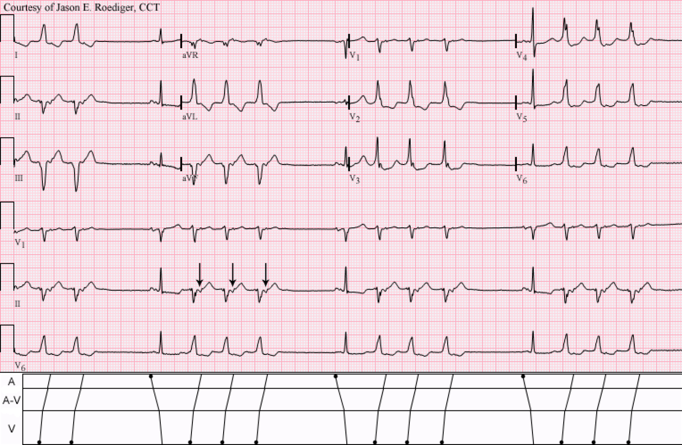
What is the rhythm seen in this 12-lead ECG?
Rate this content:
-

- jer5150's blog
- Log in or register to post comments
All our content is FREE & COPYRIGHT FREE for non-commercial use
Please be courteous and leave any watermark or author attribution on content you reproduce.


Comments
HI
Sinus prolong QT, St segments flate. Fussion beats retrograde P wave runs VT X3, coming from R side of heart. Compesentory pause might be prolong, poss. sinus Brady?
HI
Sinus prolong QT, St segments flate. Fussion beats retrograde P wave runs VT X3, coming from R side of heart. Compesentory pause might be prolong, poss. sinus Brady?
Mind the gap!
Interesting strip! Probably sinus bradycardia (mitrale P-waves in II) based on the pause duration, long QTc, diffuse ST-depression, triplet PVC's with retrograde capture of the atria. The PVC's are notable for an rS complex in V1 and a slightly shorter RP than the PR, so perhaps these are occuring close to or near the conduction tissue by the AVN.
Christopher
sixlettervariable.blogspot.com
ems12lead.com
SINUS RHYTHM
zafer karabulut
Automatic Focus
SR, SB, the baseline ECG does not show pathology except for the abnormal P wave. NSVT [3 beats] with retrograde P wave. The QRS complex of the NSVT is narrowish and looks originating close to the conduction system in the postero-septal area close to the left posterior fascicle. This is definitely automatic focus and not reentrant one!!
Awaiting Jason's Follow-Up Strip ...
Knowing Jason (at least knowing him thru his ECG expertise) - I suspect he has at least one follow-up tracing on this patient that he is planning to show us ... What I see here is underlying sinus rhythm (sinus brady) - with salvos. Each ventricular beat manifests retrograde conduction with a fixed RP. The coupling interval for the 1st PVC in each grouping is constant. Of interest - the rate for the salvo is not overly fast (~120/min) - and the R-R interval within each salvo is not precisely the same - but the pattern IS the same from one salvo to the next. So reading much more into this than I normally would (since it is a "Jason tracing" ) - I wonder about some type of exit block ...
Ken Grauer, MD www.kg-ekgpress.com [email protected]
INTERPRETATION
INTERPRETATION:
1. Sinus rhythm (? sinus bradycardia; rate indeterminate) interrrupted by . . .
2. . . . short, 3-beat trios of ventricular tachycardia with 1:1 retrograde atrial activation (arrows)
3. Intraatrial block
COMMENTS:
The computer calculated the duration of the QT interval at 390 ms and the QTc at 474 ms. There are nonspecific ST-T abnormalities. The R-P' interval on the VPBs is comparable to the conducted P-R interval on the sinus beats. When compared to other causes of "group beating", this is a relatively rare form.
It would serve no practical purpose to call this ventricular quadrigeminy. "When consecutive VPBs follow each sinus beat, we already have a better name for it since, by definition, three consecutive VPBs make the shortest definable run of tachycardia" (1)
Reference/Source:
(1) Marriott HJL. Practical Electrocardiography. 8th ed. Baltimore: William & Wilkins, 1988, p. 142
Jason E. Roediger - Certified Cardiographic Technician (CCT)
[email protected]