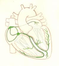
Original illustration by Dawn Altman. All content on the ECG Guru is FREE and FREE of COPYRIGHT for your use in the classroom. Please contact the ECG Guru administrator for questions about other uses. Email at [email protected]
All our content is FREE & COPYRIGHT FREE for non-commercial use
Please be courteous and leave any watermark or author attribution on content you reproduce.


Comments
emergency
How to identify atrial tachycardia on ECG?
Hasan
Atrial Tachycardia
Hi, Hasan,
To answer your question in the simplest terms, atrial tachycardia is a narrow-complex rhythm that is faster than 100 bpm, but usually between 140 and 240 bpm. The P waves are slightly different than the patient's sinus P waves, although they may be impossible to see, as they often fall in the preceding T waves. Those T waves will appear taller than the sinus T waves due to the addition of the P. The QRS will be narrow unless a separate interventricular conduction delay, like bundle branch block, occurs. Depending on the origin of the rhythm, it may be very regular with a sudden onset and offset or regular to slightly irregular with a more gradual onset
If you would like to delve deeper into the various forms of supraventricular tachycardia, search these terms: atrial tachycardia, multifocal atrial tachycardia, AVNRT, AVRT, Reentry, and paroxysmal supraventricular tachycardia. It is a very fascinating subject
Hope this helps
Dawn
Dawn Altman, Admin