
Submitted by Dr A Röschl on Mon, 09/23/2024 - 02:51
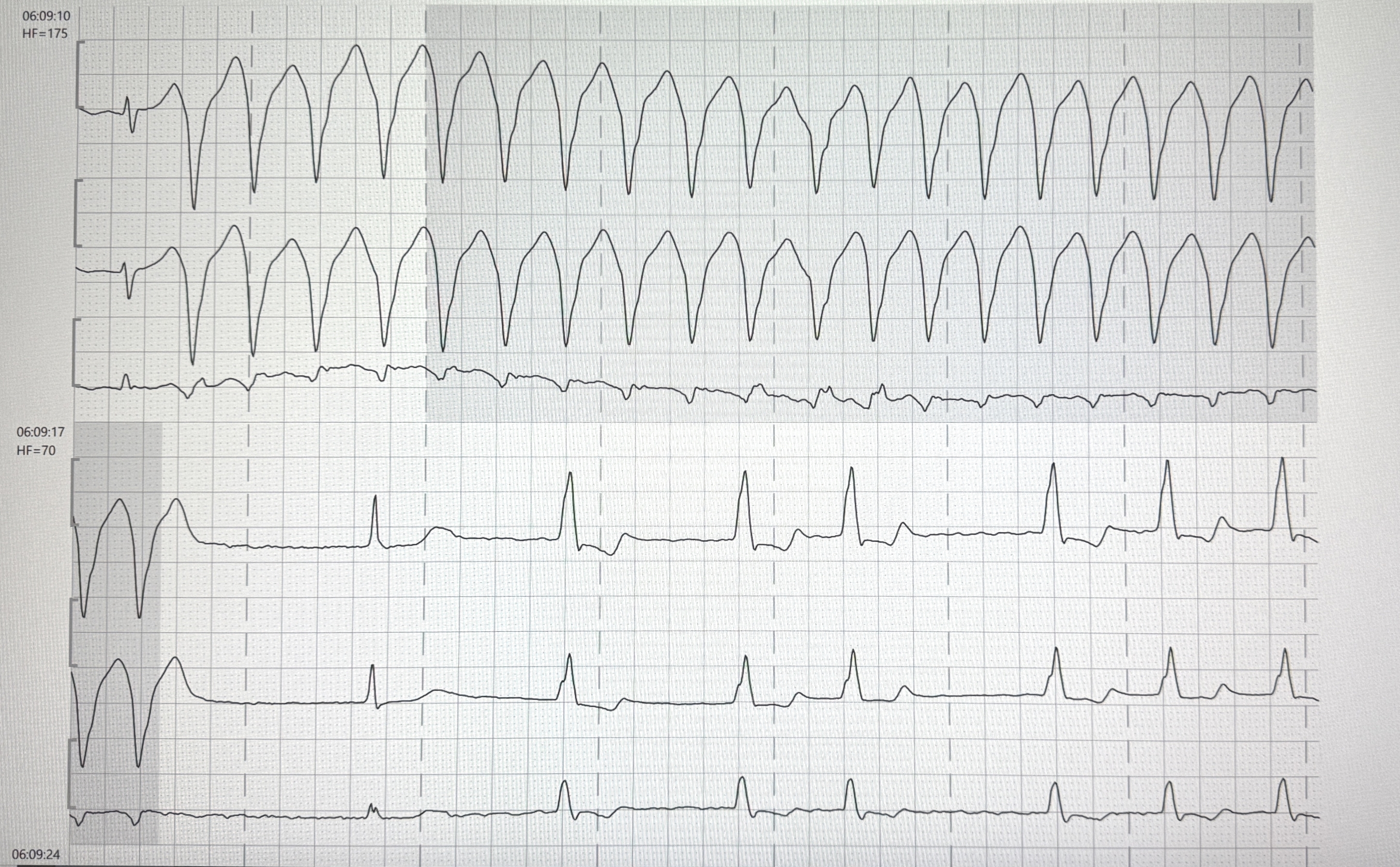
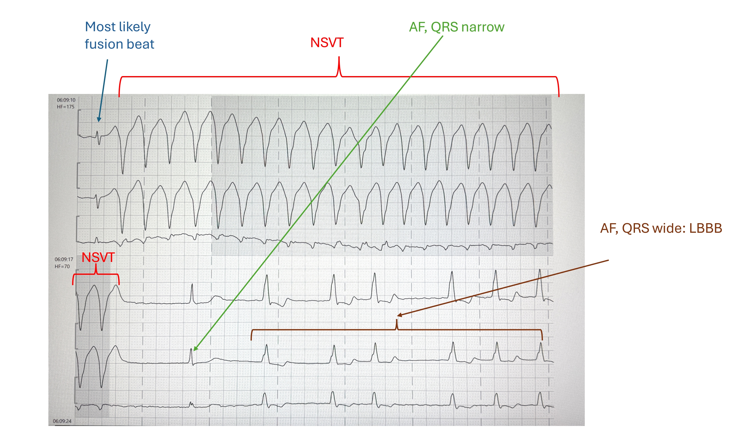
Extract from a Holter ECG, 2 continuous strips, recorded at 25 mm/s. At the top you can see a broad complex tachycardia without recognizable P waves, which ends spontaneously after 2 beats in the lower section. This is a ventricular tachycardia (VT). The very first beat in the 1st strip is most probably a fusion beat. After the end of the VT in the 2nd strip, a narrow QRS complex appears, here you can also recognize that atrial fibrillation is present. The further beats are irregular and wide, there is a bundle branch block, in this case a left bundle branch block (the initial QRS vector shows a sluggish rise). Perhaps Dawn or Ken Grauer would like to comment on this?
Rate this content:
-

- Dr A Röschl's blog
- Log in or register to post comments
All our content is FREE & COPYRIGHT FREE for non-commercial use
Please be courteous and leave any watermark or author attribution on content you reproduce.