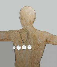
The posterior leads are placed in the fifth intercostal space with the electrode for Lead V9 placed at the left spinal border, V8 at the scapula, and V7 halfway between V6 and V8. Most commonly, the V4, V5, and V6 leadwires are used, and the printed ECG labelled to show the changes.
This is an original illustration by Dawn Altman. It may be used for no charge and free of copyright for classroom presentations. For commercial use, please contact the artist at [email protected].
All our content is FREE & COPYRIGHT FREE for non-commercial use
Please be courteous and leave any watermark or author attribution on content you reproduce.

