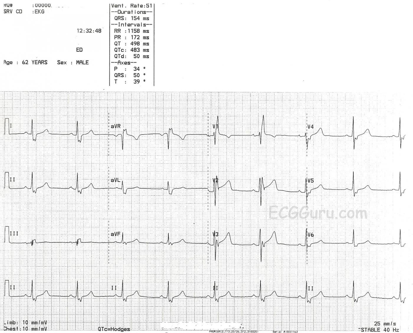
This is an example of right bundle branch block - with a couple of twists. It has the usual ECG characteristics of right bundle branch block: widened QRS (154 ms), supraventricular rhythm (sinus bradycardia), and an rSR' pattern in V1. In addition, wide little S waves are clearly seen in Leads I and V6. This secures the diagnosis of right bundle branch block (RBBB). Each QRS complex in every lead starts off with a very normal appearance, or morphology. Then, as the right ventricle is depolarized late, an additional wave is "added on". This is the R-Prime (R') in V1 and the S wave in Leads I and V6.
In most examples of RBBB, you will see the T wave point in the OPPOSITE direction of the terminal wave. So, V1 should have a NEGATIVE T wave. In this example, V2 and V3 should have also had negative T waves. The upright T waves could be considered to have the same significance as inverted T waves in a normal ECG.
Another interesting aspect to this ECG is the unusual morphology of the terminal S wave in most of the leads. There appears to be a slight notch. Lead V2 even appears to have ST elevation. Perhaps some of our Gurus would comment on this.
This is a good ECG to use to show how the terminal R' and S waves can sometimes be confused with ST elevation and depression. Lead III has a very flat T wave, and one might make the mistake of calling the R' wave "ST elevation". The R' does not have the sloping shape of a normal ST segment and T wave. Also, all the channels on the ECG are run simultaneously. One needs only to look up at Leads I and II to see where the true T waves are - Lead III's T wave is directly under them.
This is a very good teaching ECG. We look forward to hearing your comments.
All our content is FREE & COPYRIGHT FREE for non-commercial use
Please be courteous and leave any watermark or author attribution on content you reproduce.


Comments
Complete RBBB - with a few "twists"
Ken Grauer, MD www.kg-ekgpress.com [email protected]
QTc
Has anyone noticed a QT-interval prolonged?