
Submitted by Dawn on Mon, 03/12/2012 - 16:46
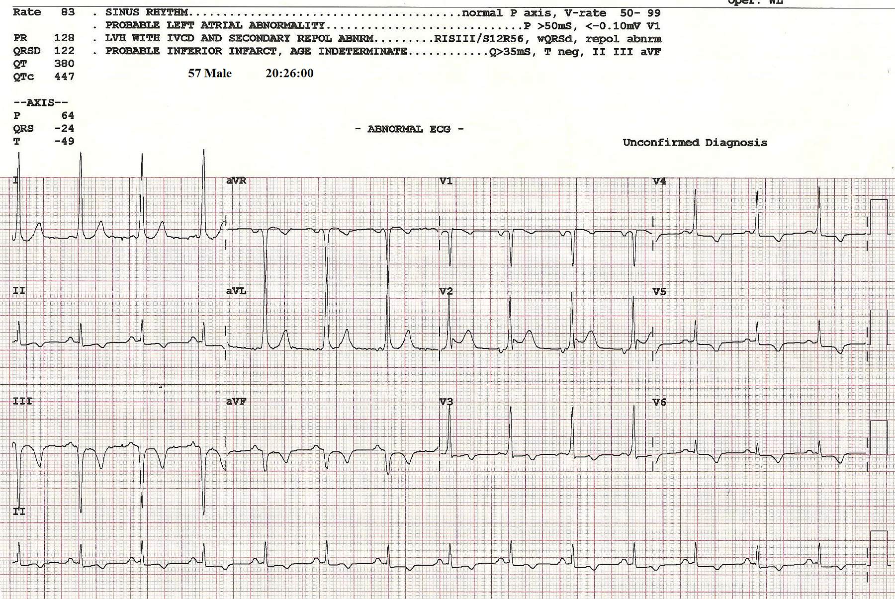
This ECG is a followup to "Wide Complex Tachycardia In a Patient with WPW". The patient was known to have Wolff-Parkinson-White syndrome, and presented to an urgent care center with a hemodynamically unstable wide complex tachycardia. He was successfully cardioverted by paramedics. This followup tracing clearly shows the Delta waves, indicating early activation of the ventricles through an accessory pathway. See Leads I, V3, and V4 for the clearest views of the Delta waves. The ST changes: ST elevation in Leads V1 and V2, and the T wave inversions and ST depressions in Leads II, III, aVF and V3 through V6, could represent an acute injury in the inferior-lateral wall, or post-cardioversion ST changes.
Thanks to ECG Guru Member, Sebastian Garay, for his contribution of this very interesting set of ECGs.
Rate this content:
All our content is FREE & COPYRIGHT FREE for non-commercial use
Please be courteous and leave any watermark or author attribution on content you reproduce.