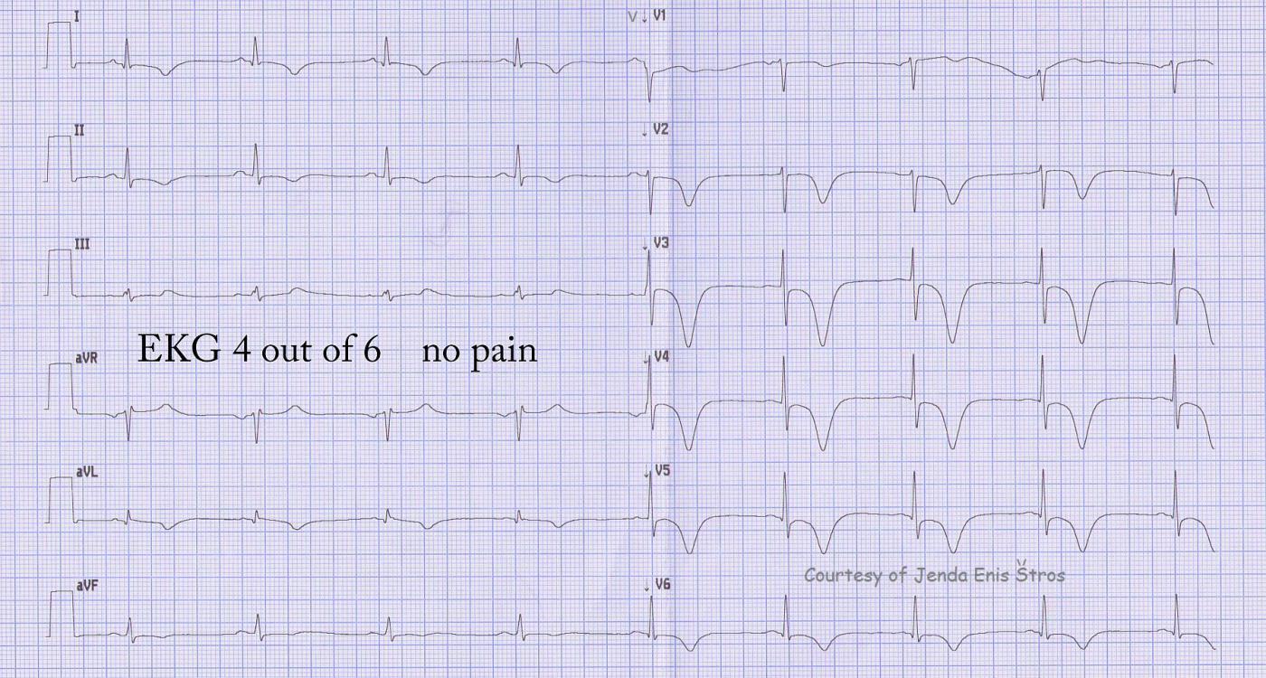
Continuing our teaching series of ECGs donated by Jenda Enis Štros, ECG 4 of 6 shows a new occurance of huge T wave inversions in the precordial leads. Since this is the area that was stented (left anterior descending artery, anterior wall of the LV), we immediately should think of re-occlusion of the artery. In a newly-placed stent, the danger is thrombosis (blood clot). The patient had no chest pain at this time.
Here are links to all six of the ECGs in this series:
http://www.ecgguru.com/ecg/teaching-series-1113-ecg-1-6-acute-anterior-wall-mi
http://www.ecgguru.com/ecg/teaching-series-1113-ecg-2-6-acute-anterior-wall-mi
http://www.ecgguru.com/ecg/teaching-series-1113-ecg-3-6-acute-anterior-wall-mi
http://www.ecgguru.com/ecg/teaching-series-1113-ecg-4-6-acute-anterior-wall-mi
http://www.ecgguru.com/ecg/teaching-series-1113-ecg-5-6-acute-anterior-wall-mi
http://www.ecgguru.com/ecg/teaching-series-1113-ecg-6-6-acute-anterior-wall-mi
All our content is FREE & COPYRIGHT FREE for non-commercial use
Please be courteous and leave any watermark or author attribution on content you reproduce.


