Dawn's picture

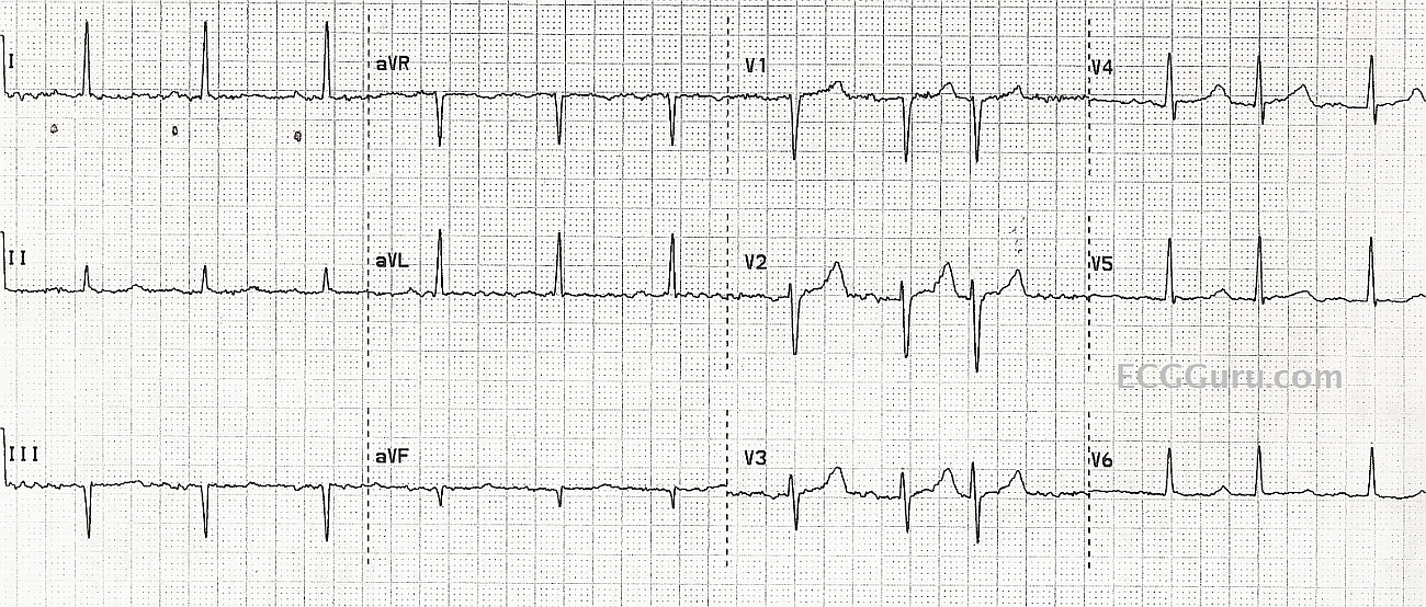
This patient was diagnosed by the rescue crew as having atrial fibrillation, based on the fact that they thought the rhythm was irregular, and they could not see P waves.  They also noted a wavy baseline, and considered it to be fibrillatory waves.  In reality, the underlying rhythm is regular, with some PACs (regularly irregular). The P waves are small and hard to see in the baseline artifact.  We have marked the P waves in Lead I with small dots.  

It pays to look at multiple leads, reduce artifact as much as possible, and look at the strip for evidence of an underlying rhythm.  

It is not shown here, but the ECG machine is often able to show that the P waves are present by giving a PR interval and P wave axis in the diagnostics.

Rate this content: 
Average: 4 (5 votes)

Comments

ekgpress@mac.com's picture

 
This is a TOUGH tracing! — and IF I was the provider, I would NOT be 100% certain that the rhythm is sinus with PACs from this single 12-lead ECG. The problem is that we are not given a simultaneously-recorded long lead II rhythm strip (as pre-hospital tracings often are missing a long lead rhythm strip, and only have 12 leads). Some concepts are worth reviewing:
  • There are 12 “beats” on this tracing. For each of these 12 beats — we get “3 looks” (in leads I,II,III; aVR,L,F; V1,2,3; and V4,5,6). Sinus rhythm is defined by the presence or absence of an upright P wave in lead II — and we unfortunately ONLY have 3 beats in lead II to look at. Of these, the deflection preceding beat #1 is too small and deformed to call it sinus …. It DOES look like we DO have a very small but upright P wave with fixed PR interval preceding beats #2 and 3 — but I am NOT convinced from looking at the other 9 leads that a similar morphology P wave with constant PR interval is present. IF we had a long lead II with 12 beats in lead II — I’m virtually certain we would be able to tell IF what seems to be present preceding beats #2 and #3 is real or not … but we don’t!
  • I measured R-R intervals with calipers. Although the R-R interval is VERY similar for the first 8 beats — it is NOT identical! And the R-R interval is clearly variable for the remaining 4 beats. So while I would SUSPECT the rhythm is SINUS from looking at this 12-lead, because of the above-described appearance of beats #2 and 3 in lead II, and because of the almost identical R-R intervals for these first 8 beats — the tiny size of P waves and the baseline artifact plus indefinite and variable P wave appearance in the other 9 leads leave me UNCERTAIN of the diagnosis …
BOTTOM LINE: Additional monitoring is needed to definitively say what the rhythm is in this case — though I suspect it is sinus with PACs.
--------------------------------------------------------
  • NOTE: I have developed a FREE On-Line ECG Video Series on the Basics of Rhythm Interpretation. The 3 parts (my Video Blogs #10,11,12) in this series start from the very beginning, with plenty of PEARLS sprinkled along the way for intermediate and advanced interpreters. The link I give is of a detailed CONTENTS that allows you to immediately jump to ANY subject within this 2 Hour-long series. 
 

Ken Grauer, MD  www.kg-ekgpress.com   [email protected] 

Dawn's picture

thanks so much, Dr. Ken. I should have added the information that the subsequent ECG in the Emergency Dept. Clearly showed sinus rhythm with occasional PACs. That tracing has been misplaced, unfortunately. I hoped to make a point that a sub-optimal ECG, with artifact, makes it nearly impossible to make an accurate interpretation. 


Dawn Altman, Admin

All our content is FREE & COPYRIGHT FREE for non-commercial use

Please be courteous and leave any watermark or author attribution on content you reproduce.