Submitted by Dawn on Mon, 08/13/2012 - 10:51
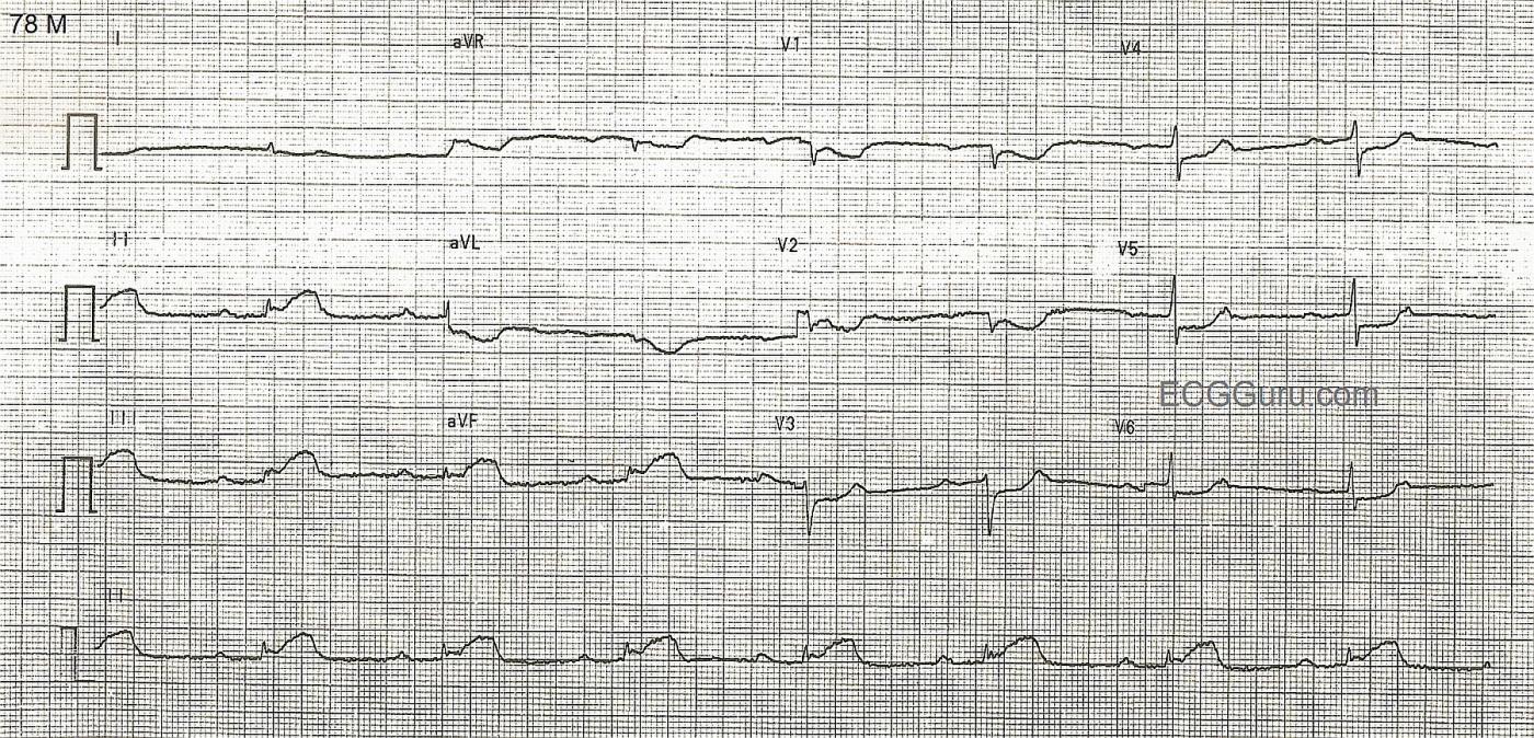
Inferior wall MI: ST elevation in II, III, and aVF. Reciprocal ST depressions. Sinus bradycardia and first-degree AV block suggests sinus node and AV node ischemia. This is a good "classic" inferior wall M.I. It is good for teaching inferior-posterior injury, and the effects of RCA occlusion on the sinus and AV nodes. The low voltage in the limb leads may also be due to acute M.I., but in this case, we do not know the patient's body size.
Related Terms:
Rate this content:
All our content is FREE & COPYRIGHT FREE for non-commercial use
Please be courteous and leave any watermark or author attribution on content you reproduce.


Comments
Mechanisms of Bradycardia During AMI
Thank you for the nice tracing. Of course sinus bradycardia is much more common in Inferio-posterior MI than other infarctions. SB can be caused by ischemic to the SA node, which is supplied by an atrial branch of the proximal RCA in 55% and from proximal Cx Artery in 45%. However, the presence of prolongation of PR interval idicates ischemia at the level of AV node, which is mostly supplied by RCA.
The other mechanisms of SB are: 1. Vagally mediated, 2. Bezold Jarisch Reflex, 3. Atrial Infarction, 4. Metabolic [Hyperkalemia,. . . etc.]. In this nice example, ther is no evidence of atrial infarction.
Final comment, the combination of SB and AV delay [first degree AV block] is caused by ischemia but can be vagally mediated without ischemia of the SAN or the AVN.
Thank you
Thank you Dr Sham'a for this complete and concise explanation. Your contribution to this content will be a great help to instructors and other ECG Gurus.
Dawn Altman, Admin