Dawn's picture

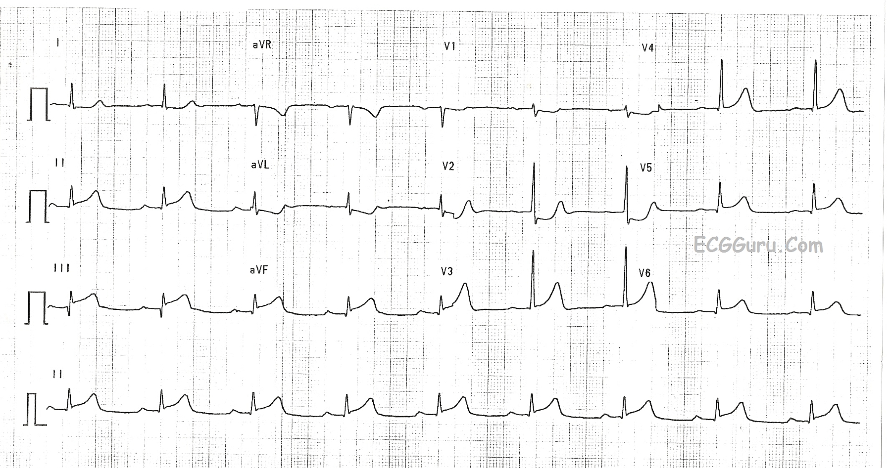
This ECG shows a classic inferior - posterior STEMI.  This M.I. was due to complete occlusion of the right coronary artery.  ST elevation apparent in Leads II, III, and aVF show the acute injury in the inferior wall, while ST depressions in V1 and V2 are reciprocal of the ST elevations in the posterior wall.  The tall R waves in Leads V1 - V3 most likely are reciprocal to pathological Q waves in the posterior wall.  Tall R waves in the right precordial leads can be caused by other cardiac conditions, such as right ventricular enlargement.  RV hypertrophy can probably be ruled out in this case because there is no right axis deviation or P pulmonale.  Because inferior wall M.I.s often extend into the posterior wall, it is the most likely cause of the tall R waves.

Rate this content: 
Average: 4.4 (7 votes)

Comments

ekgpress@mac.com's picture

Excellent illustrative example by Dawn of acute infero-postero MI. I'd add the following points to her explanation:

  • The RCA (Right Coronary Artery) - generally supplies the right ventricle and posterior and inferior walls of the left ventricle. This explains why inferior & posterior MI's are commonly seen together.
  • While right-sided precordial leads would be needed to know for sure about acute RV infarction - I'd be less suspicious of this in this case (unless the patient was hypotensive or became hypotensive after NTG) because there is ST depression in lead V1. Often you'll see either a flat or slightly elevated ST segment in V1 (but not in V2,V3) when there is associated RV involvement.
  • In theory - acute infero-postero MI could be due to either: i) acute RCA occlusion; or ii) acute circumflex (LCx) occlusion in a dominant-left circulation (in which the RCA is a smaller less dominant vessel). Approximately 80-90% of individuals have a right-dominant circulation - so acute RCA occlusion is far more common.
  • Clues in this tracing that the lesion is in the RCA are: i) ST elevation in lead III>II; and ii) marked ST depression in lead aVL >lead I.
  • Note that in addition - there are small q waves in V4,V5,V6 - and ST elevation (!) also in leads V5,V6. Although one might be tempted to suspect a left-dominant circulation (due to infero-poster-"lateral") involvement - LEAD V6 ST elevation may also be a "reciprocal change" from posterior infarction (and it is not necessarily due to "lateral" infarction). Given how much more marked ST elevation is in lead III>II - I'd suspect RCA occlusion in this case (confirmed by Dawn's revelation that RCA occlusion was in fact the "culprit" artery).
  • Practically speaking - it doesn't matter all that much what the "culprit artery" will be (as fun as it is to predict) - since it is obvious from this ECG that the patient needs acute cath - and definitive answers will then be obtained (sometimes collateralization and anatomic variants may account for unusual patterns).
  • Finally - as Dawn suggests - there IS an important differential diagnosis to keep in mind for the "Tall R wave in lead V1". I totally agree in this case that the disproportionately tall R waves in V2,V3 are undoubtedly the result of acute posterior MI given the changes described above. Moreover, the R wave in V1 is really not taller than the S wave - so this example is really not consistent with things like RVH, RBBB, etc.
  • Additional educational material that may be of interest include: i) Brief pdf on the "Mirror Test" (which is positive in lead V2 of this tracing!) and which also contains the List of Causes for a Tall R in Lead V1 - and ii) Review of How to PREDICT the CULPRIT ARTERY.

Ken Grauer, MD  www.kg-ekgpress.com   [email protected] 

All our content is FREE & COPYRIGHT FREE for non-commercial use

Please be courteous and leave any watermark or author attribution on content you reproduce.