
Submitted by Dawn on Tue, 04/28/2015 - 20:22
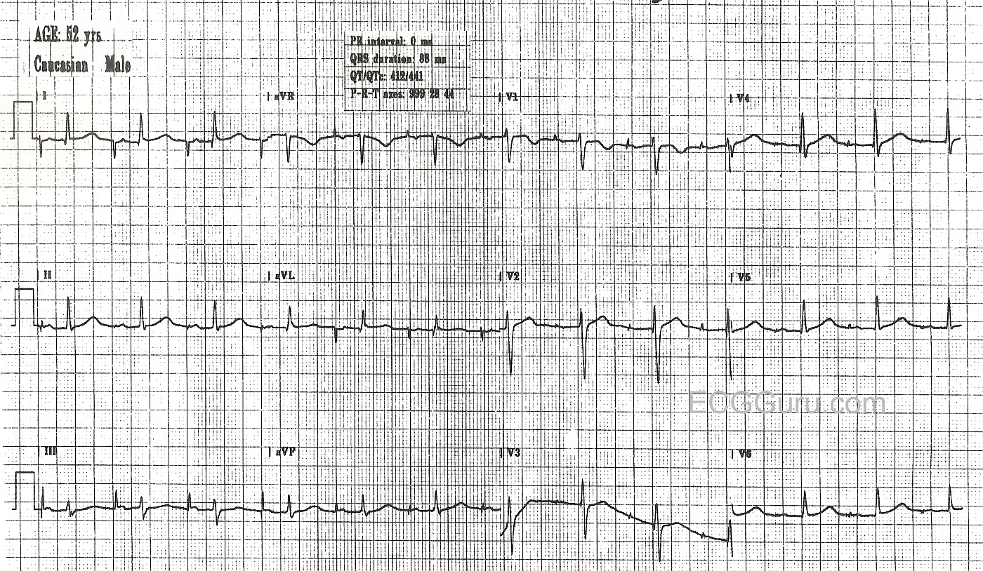
This is a good example of an AV Sequential pacemaker in a patient with an intact AV conduction system. The pacemaker is pacing the right atrium, and the impulse is being transmitted normally down through the AV node and the interventricular conduction system. The pacer spike is seen before the P waves, and the QRS complex is narrow, reflecting normal conduction through the ventricles.
If you are teaching about ST elevation MI, this patient has no ST elevation M.I., but this type of pacing does not affect the ST segments, and an M.I. will still show as ST elevation.
Related Terms:
Rate this content:
All our content is FREE & COPYRIGHT FREE for non-commercial use
Please be courteous and leave any watermark or author attribution on content you reproduce.
Comments
Atrial Pacing (with intermittent Ventricular Spikes)
As per Dawn - this ECG provides a nice example of atrial pacing. As always with any pacemaker — it is essential to know what parameters have been set in order to interpret the tracing. It looks like the PR interval (duration from onset of atrial spike until onset of the QRS) = 0.24 second. Depending on the patient’s underlying disorder — the programming cardiologist can set parameters to optimize the chance for spontaneous initiation of ventricular contraction if the conduction system is intact.
Ken Grauer, MD www.kg-ekgpress.com [email protected]