Dawn's picture

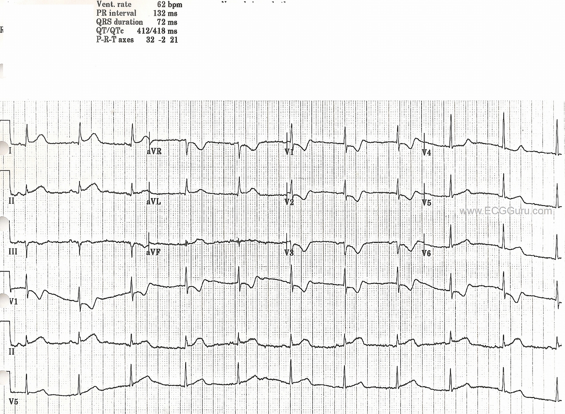
This ECG is from old files, and no patient information is available.  Acute ST elevation is apparent in lateral leads I and aVL, and in Lead II.  In addition, the anterior leads V1, V2, and V3 show reciprocal ST depression and, in V1 and V2, taller-than-normal R waves.  This points to acute posterior - lateral M.I., probably due to occlusion of the circumflex artery. Unfortunately, we do not have angiogram confirmation for this.

The P waves are interesting.  They are hard to see because of their small size and the fine baseline artifact present.  The best view appears to be in the Lead II rhythm strip at the bottom.  In this view, the P waves' morphology appears to be changing.  What do you think?


Related Terms: 
Rate this content: 
Average: 3.4 (5 votes)

Comments

ekgpress@mac.com's picture

This tracing is as good as it gets for teaching. Dawn alludes to many of the interesting points. I'll add the following observations:

  • The rhythm is uncertain. There is just too much baseline artifact to be certain. Recognizing that in itself is a helpful teaching point. The R-R interval is fairly (although not precisely) regular. I think the rhythm is sinus - because it looks like the P wave is upright in lead II. But if all I had was a lead II rhythm strip - I would not be at all certain. This is the value of having a full 12-lead. Support that sinus rhythm is the likely mechanism is forthcoming from some "unlikely" sources - namely what looks to be a reasonably consistent atrial deflection with constant PR interval in leads I, aVR and V3.
  • Another reason to really try and hone in on the rhythm is to be sure we aren't missing 2:1 AV block. As we'll discuss in a moment - there is obvious acute infarction. Given the relatively slower rate and uncertain P wave etiology - the question of whether this might be 2:1 should be entertained. Turns out it isn't - but before I was comfortable with that assertion, I had to set my calipers to precisely half the R-R interval to look and see if extra P waves could have been present. They are not. So my bet is sinus rhythm at 60-65/minute - though clinically I'd want to confirm that by review of additional tracings.
  • Otherwise - this tracing is as good of an example of presumed acute Circumflex occlusion with resultant acute postero-lateral infarction as you are likely to see. Note the disproportionately Tall R Wave in lead V1 with positive "Mirror Test" in leads V1,V2,V3 (Click HERE for Review of these 2 key concepts). If you were to print out a hard copy of this tracing - then flip it over and hold it up to the light  - you'll see the "positive mirror test" (in which the tall R and shape of ST-T depression in the anterior leads looks like a large Q and ST elevation in this "mirror image" which reflects what is going on in the posterior wall).
  • Most (85-90%) of the time - acute posterior MI is from acute RCA (right coronary artery) occlusion. This is NOT the case here. Instead of inferior ST elevation - we have high lateral ST elevation in leads I and aVL. Note in addition the hyperacute morphology of the ST segment in these leads - which of itself is the mirror image of the ST-T depression that we see in V1,V2,V3. So this patient almost certainly has a dominant left circulation with occlusion of that large circumflex vessel.
  • There are small and narrow q waves in leads I and aVL. We have no way of knowing (without a prior tracing) if these are new (from acute high lateral infarction) or simply reflect normal septal q waves that are so commonly seen in lateral leads. It really doesn't matter in view of all else that is happening.

Ken Grauer, MD  www.kg-ekgpress.com   [email protected] 

I'm late to the game here (reviewing some PWMI cases I have saved) but I just noticed that it looks like V1 and V3 were probably swapped. It accounts for the strange R-wave progression and also the presence of such well-defined p-waves in "V3."

Dawn's picture

Agreed!

Dawn Altman, Admin

All our content is FREE & COPYRIGHT FREE for non-commercial use

Please be courteous and leave any watermark or author attribution on content you reproduce.