
Submitted by Dawn on Fri, 02/24/2012 - 21:20
Resource File:
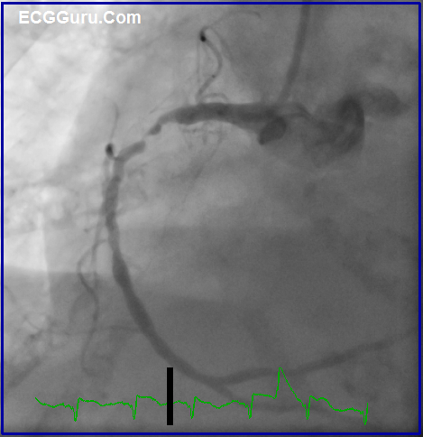
The is the right coronary artery from patient AW103, who suffered an antero-lateral M.I. after apparently suffering an inferior wall M.I. a few days previously. The right coronary artery showed signs of being completely occluded, then reperfusing as the clot broke down. Unfortunately, there was akinesis in the inferior wall, as well as in the anterior wall where the newer M.I. was located. The patient unfortunately did not survive this injury. See ECG AW103 and two other images in the Resources files.
Category:
All our content is FREE & COPYRIGHT FREE for non-commercial use
Please be courteous and leave any watermark or author attribution on content you reproduce.