Submitted by jer5150 on Sun, 09/09/2012 - 19:02
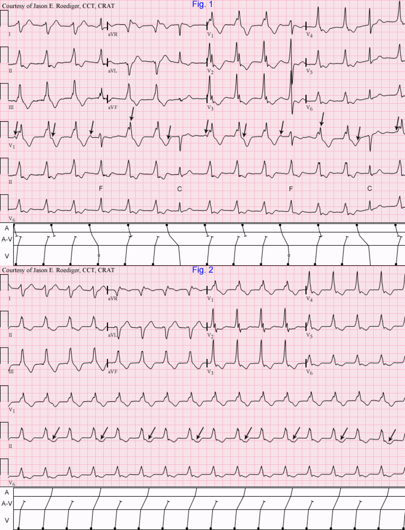
Patient's clinical data: 76-year-old white man admitted to the ICU.
Hint: In Fig. 2, there is an extremely subtle clue on that ECG that I almost didn't notice. Laddergrams will be provided for both of these as the end of the week.
What is going on here?
Rate this content:
-

- jer5150's blog
- Log in or register to post comments
All our content is FREE & COPYRIGHT FREE for non-commercial use
Please be courteous and leave any watermark or author attribution on content you reproduce.


Comments
INTERPRETATION
Fig. 1: Accelerated idioventricular rhythm (AIVR) at a rate of about 88/min dissociated from a sinus rhythm (rate about 62/min). The ectopic ventricular beats display a right axis deviation (RAD) and positive (+) concordance in the precordial leads. The 4th and 11th beats are ventricular fusions (F) and the 7th and 14th beats are ventricular captures (C). When the patient was in sinus rhythm, their QRS morphology was identical to that of the capture beats. This is A-V dissociation due to usurpation and not default. The retrograde concealed conduction into the A-V node that lengthens the P-R interval on both the fusion and capture beats. (see A-V tier in laddergrams). Presumably, this is originating in the left ventricle (LV).Fig. 2: Accelerated ventricular rhythm (rate about 99/min) with 2:1 retrograde ventriculoatrial (V-A) conduction to the atria. This is confirmed by the deformity to the T-wave on alternate beats and a constant R-P interval. These ectopic ventricular impulses are "capturing" the atria. Since the atria are under the direct control of the ventricles, one could technically drop the prefix "idio" and just call it accelerated ventricular rhythm. (see laddergram). Note that in Fig. 1, the atria remain solely under the control of the sinus node but in Fig. 2 the sinus node is suppressed by the constant retrograde atrial activation. In both tracings, the QRS duration measures "wide-wide" between 0.16 and 0.18s. Right axis deviation (RAD). Positive concordance.
Jason E. Roediger - Certified Cardiographic Technician (CCT)
[email protected]