Dawn's picture

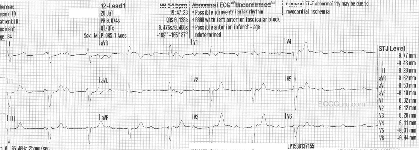
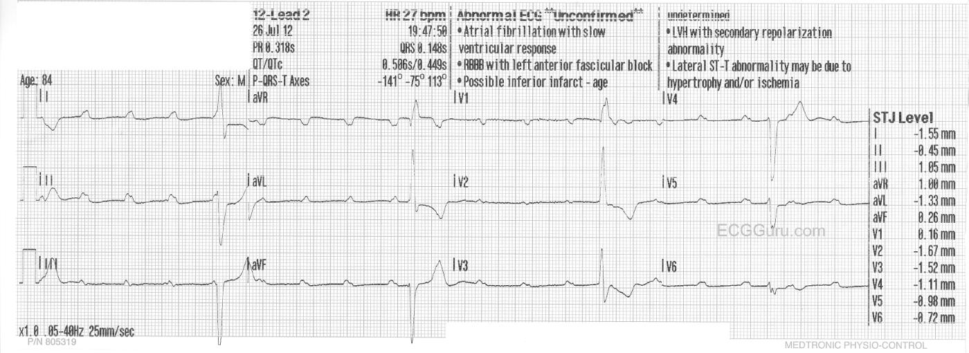
This week's ECG of the WEEK was donated to us by Sebastian Garay. These two ECGs were obtained less than 30 seconds apart from an 84 year-old man who called fire-rescue because he felt dizzy and fell.  He was not injured in the fall, and his vital signs remained stable, with an adequate BP.  These two ECGs were obtained prior to arrival in the Emergency Dept.

The first one shows a sinus rhythm at about 110/min.  There is a complete heart block (third-degree AV block), and the escape rhythm is a wide-complex rhythm at a rate of about 54/min and slowing severely toward the end.  The second ECG was taken less than 30 seconds after the first, and shows a significantly slower escape rhythm rate at 27/min., while the sinus rate increases to 120/min.  The change is sinus rate is likely an attempt by the nervous system to compensate for the lower cardiac output as the ventricular rate slows. The escape rhythm is not only slower, but there are some changes in the QRS morphology from the first ECG.

For your basic students, this ECG serves to demonstrate the AV dissociation seen in complete heart block.  It is easy to "march out" the P waves, and see that some of them are "hiding" in the QRS comlexes.  It also shows how quickly a rhythm can change rates.

For your more advanced students, you will want to have a discussion about escape rhythms.  This one initially has a fairly fast rate, suggesting junctional origin. The QRS morphology is of the right bundle branch type, with left anterior fascicular block.  However, ventricular rhythms originating from the posterior fascicle region can have the "RBBB / LAFB" morphology.  If this escape rhythm is fascicular (ventricular) in origin, it is an accelerated idioventricular rhythm.  The second escape rhythm appears very similar to the first, with the very noticeable exceptions of QRS morphology, especially in V1 and V2, and the rate.

This patient was given Atropine in the ED, with no change to the rhythm.  We do not know what transpired after that, but suspect a pacemaker was in his future. 

We look forward to comments from our members about these two very interesting ECGs.


Rate this content: 
Average: 5 (3 votes)

Comments

ekgpress@mac.com's picture

This is a highly interesting set of tracings (Sebastian's contributions always are! ) - with important teaching/learning points for ALL level providers. I'll make the following observations to add to Dawn's introductory comments:

  • Unfortunately - there is no long lead II rhythm strip for either tracing. NO MATTER - since we can still interpret the basics of these 2 rhythms (though for some subtle points, such as determining if there is high-grade vs complete AV block - it really would have been nice to see a long lead II! ).
  • These 2 ECGs are wonderful examples of high-grade-to-complete AV block tracings in which you can practice "walking out P waves". Calipers are essential for doing this.
  • KEY in the above exercise is to pay attention to the atrial rate. As Dawn mentions - the atrial rate is FASTER in the 2nd tracing (done at 19:47:50 - or 27 seconds after the 1st ECG done at 19:47:23). The atrial rate doesn't matter so much when there is true complete AV block - since NO atrial impulses are able to conduct to the ventricles, regardless of the atrial rate. BUT - it is important to be aware that sometimes with 2nd degree (or with high-grade) AV block - that a change in the ATRIAL rate may alter the number of impulses that are able to be conducted to the ventricles. Thus, it may look like the block is "getting worse" (ie, if the ventricular rate slows) - when in reality, there has been NO CHANGE in the "severity" of AV block - but simply a change in the atrial rate. For example - at an atrial rate of 80/minute - the AV node may be able to conduct 1:1 (in which case the ventricular rate will be 80/minute) - BUT - if the atrial rate speeds up to 100/minute, the diseased AV node may only be able to conduct 1:2 at this faster atrial rate - which might therefore result in an overall slower ventricuar rate of 100/2 = 50/minute.
  • CLINICALLY - this sequence of events may occur when ATROPINE is given. Atropine may improve AV conduction - but it also tends to speed up the atrial rate. This explains the sometimes paradoxical "slowing effect" when atropine is given to advanced AV blocks (ie, by speeding up the atrial rate - the overall ventricular rate may paradoxically slow .... ). So Atropine may help some patients - but it DOES also have potential to harm .... which is why the drug is best utilized (in an "ideal" world) for AV blocks with a narrow QRS during the early hours of acute inferior infarction when increased vagal tone is most likely to be the "culprit" for causing the AV block ....
  • Although possible that there is junctional escape in the TOP (1st) tracing - I would call this AIVR (Accelerated IdioVentricular Rhythm) at 54/minute until proven otherwise. It would seem that an only slightly "accelerated" ventricular escape rhythm with a very wide and bizarre QRS is far more likely to represent what we see here than escape from the AV node ...
  • Did you catch the sustained PAUSE at the end of the TOP tracing? Since the ECG ends - We have NO IDEA if this is the onset of asystole or simply a brief period of ventricular standstill ..... Presumably it is the latter (ie, a period of ventricular standstill) - which explains why a DIFFERENT ventricular escape focus (this time a slower one) has to arise on the BOTTOM TRACING (otherwise the patient would be asystolic). So we now KNOW the reason for the difference in QRS morphology (that Dawn mentioned) between TOP and BOTTOM tracings - since we now have 2 DIFFERENT ventricular escape foci, the 2nd being much slower .... Bottom Line - A PACER IS NEEDED ASAP!
  • The BOTTOM TRACING might not be complete AV block !!!!  This is where I'd LOVE to have a simultaneous long lead II (to know for sure) - but note how precise the R-R interval is for the first 3 beats (11.4 large boxes) - yet it is SHORTER for the 4th beat in the BOTTOM tracing (~10 large boxes). Although it is possible to have complete AV block with an irregular ventricular rate - I think it is MUCH MORE LIKELY that the BOTTOM tracing represents high-grade AV block with conduction of the last impulse (I might think different if the R-R for the other beats was not so precisely regular). That said - clinically, it matters not - since regardless of whether this represents very high grade AV block with a very slow and wide ventricular escape pacemaker or complete AV block - a PACER IS NEEDED ASAP. That said - for your advanced students (to keep them honest and learning) - I'd make the point that they should recognize that the R-R interval for the last beat in the BOTTOM TRACING is shorter - and that this MIGHT mean that the degree of AV block was not complete at this moment ....

GREAT set of tracings!

  • For anyone interested - Click on this Link to my Web Page on the Basics of AV Block (contains links to free downloads on AV block, plus links to a bunch of tracings with detailed explained answers).

Ken Grauer, MD  www.kg-ekgpress.com   [email protected] 

All our content is FREE & COPYRIGHT FREE for non-commercial use

Please be courteous and leave any watermark or author attribution on content you reproduce.