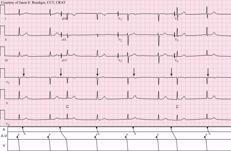
This patient was seen by his primary care provider (PCP) on an outpatient basis. The PCP decided to send her patient over to me to perform a routine ECG and establish a baseline, hince the computer's statement below of "No previous ECGs available". I printed out the above 12-lead ECG and became slightly concerned with the rhythm I was seeing. Consequently, I also recorded six full pages of continuous rhythm (not shown here). I don’t ordinarily resort to doing this
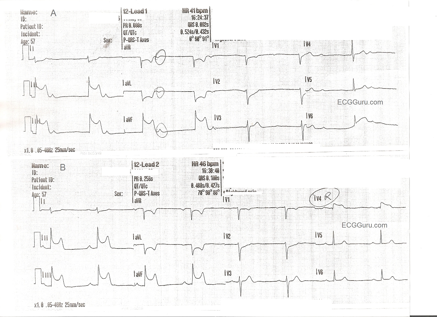
These two ECGs are from a 57 year old man with chest pain. The initial ECG shows ST elevation in Leads II, III, and aVF - inferior wall STEMI. Reciprocal changes are as expected in I and aVL. Reciprocal ST depression also seen in V1 and V2 indicate probable posterior wall involvement. Not surprising since the inferior wall is simply the lower part of the posterior wall. The first ECG also shows the patient in sinus brady with junctional escape: AV dissociation. The sinus node is often affected in IWMI that is caused by right coronary artery occlusion. The second ECG shows a slight increase in the sinus rate, and a sinus bradycardia. A V4 right lead has been performed, clearly showing ST elevation, and indicating right ventricular M.I.
FOR MORE INFORMATION ABOUT RIGHT VENTRICULAR M.I.,